- 靶点:
- HSP27
- 简介:
- >>MAPK signaling pathway;>>VEGF signaling pathway;>>Amoebiasis
- 基因名称:
- HSPB1
- 蛋白名称:
- Heat shock protein beta-1
- Human Gene Id:
- 3315
- Human Swiss Prot No:
- P04792
- Mouse Gene Id:
- 15507
- Mouse Swiss Prot No:
- P14602
- Rat Gene Id:
- 24471
- Rat Swiss Prot No:
- P42930
- 免疫原:
- The antiserum was produced against synthesized peptide derived from human HSP27. AA range:48-97
- 特异性:
- HSP27 Polyclonal Antibody detects endogenous levels of HSP27 protein.
- 组成:
- Liquid in PBS containing 50% glycerol, 0.5% BSA and 0.02% sodium azide.
- 来源:
- Polyclonal, Rabbit,IgG
- 稀释:
- IF 1:50-200 WB 1:500 - 1:2000. IHC 1:100 - 1:300. ELISA: 1:20000. Not yet tested in other applications.
- 纯化工艺:
- The antibody was affinity-purified from rabbit antiserum by affinity-chromatography using epitope-specific immunogen.
- 浓度:
- 1 mg/ml
- 储存:
- -15°C to -25°C/1 year(Do not lower than -25°C)
- 其他名称:
- HSPB1;HSP27;HSP28;Heat shock protein beta-1;HspB1;28 kDa heat shock protein;Estrogen-regulated 24 kDa protein;Heat shock 27 kDa protein;HSP 27;Stress-responsive protein 27;SRP27
- 实测条带:
- 27kD
- 背景:
- The protein encoded by this gene is induced by environmental stress and developmental changes. The encoded protein is involved in stress resistance and actin organization and translocates from the cytoplasm to the nucleus upon stress induction. Defects in this gene are a cause of Charcot-Marie-Tooth disease type 2F (CMT2F) and distal hereditary motor neuropathy (dHMN). [provided by RefSeq, Oct 2008],
- 功能:
- disease:Defects in HSPB1 are a cause of distal hereditary motor neuronopathy type 2B (HMN2B) [MIM:608634]. Distal hereditary motor neuronopathies constitute a heterogeneous group of neuromuscular disorders caused by selective impairment of motor neurons in the anterior horn of the spinal cord, without sensory deficit in the posterior horn. The overall clinical picture consists of a classical distal muscular atrophy syndrome in the legs without clinical sensory loss. The disease starts with weakness and wasting of distal muscles of the anterior tibial and peroneal compartments of the legs. Later on, weakness and atrophy may expand to the proximal muscles of the lower limbs and/or to the distal upper limbs.,disease:Defects in HSPB1 are the cause of Charcot-Marie-Tooth disease type 2F (CMT2F) [MIM:606595]. CMT2F is a form of Charcot-Marie-Tooth disease, the most common inherited disorder of
- 细胞定位:
- Cytoplasm . Nucleus . Cytoplasm, cytoskeleton, spindle . Cytoplasmic in interphase cells. Colocalizes with mitotic spindles in mitotic cells. Translocates to the nucleus during heat shock and resides in sub-nuclear structures known as SC35 speckles or nuclear splicing speckles. .
- 组织表达:
- Detected in all tissues tested: skeletal muscle, heart, aorta, large intestine, small intestine, stomach, esophagus, bladder, adrenal gland, thyroid, pancreas, testis, adipose tissue, kidney, liver, spleen, cerebral cortex, blood serum and cerebrospinal fluid. Highest levels are found in the heart and in tissues composed of striated and smooth muscle.
Identification and Comparison of Differentiation-Related Proteins in Hepatocellular Carcinoma Tissues by Proteomics:. TECHNOLOGY IN CANCER RESEARCH & TREATMENT 2017 Sep 25 IHC Human Hepatocellular carcinoma (HCC) tissue
货号:YT2252
Extracellular vesicles derived from monomeric α-synuclein-treated microglia ameliorate neuroinflammation by delivery of miRNAs targeting PRAK NEUROSCIENCE LETTERS Na Li WB Mouse 1:1000 BV2 microglia
货号:YT2252
- June 19-2018
- WESTERN IMMUNOBLOTTING PROTOCOL
- June 19-2018
- IMMUNOHISTOCHEMISTRY-PARAFFIN PROTOCOL
- June 19-2018
- IMMUNOFLUORESCENCE PROTOCOL
- September 08-2020
- FLOW-CYTOMEYRT-PROTOCOL
- May 20-2022
- Cell-Based ELISA│解您多样本WB检测之困扰
- July 13-2018
- CELL-BASED-ELISA-PROTOCOL-FOR-ACETYL-PROTEIN
- July 13-2018
- CELL-BASED-ELISA-PROTOCOL-FOR-PHOSPHO-PROTEIN
- July 13-2018
- Antibody-FAQs
- 产品图片
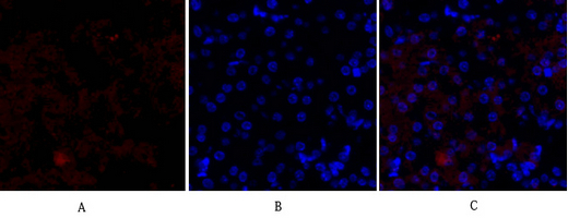
- Immunofluorescence analysis of mouse-kidney tissue. 1,HSP27 Polyclonal Antibody(red) was diluted at 1:200(4°C,overnight). 2, Cy3 labled Secondary antibody was diluted at 1:300(room temperature, 50min).3, Picture B: DAPI(blue) 10min. Picture A:Target. Picture B: DAPI. Picture C: merge of A+B
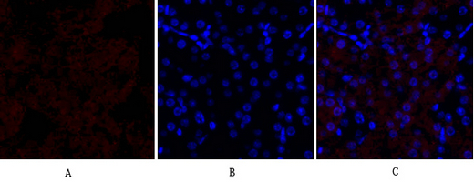
- Immunofluorescence analysis of mouse-kidney tissue. 1,HSP27 Polyclonal Antibody(red) was diluted at 1:200(4°C,overnight). 2, Cy3 labled Secondary antibody was diluted at 1:300(room temperature, 50min).3, Picture B: DAPI(blue) 10min. Picture A:Target. Picture B: DAPI. Picture C: merge of A+B

- Immunohistochemical analysis of paraffin-embedded Human-stomach-cancer tissue. 1,HSP27 Polyclonal Antibody was diluted at 1:200(4°C,overnight). 2, Sodium citrate pH 6.0 was used for antibody retrieval(>98°C,20min). 3,Secondary antibody was diluted at 1:200(room tempeRature, 30min). Negative control was used by secondary antibody only.
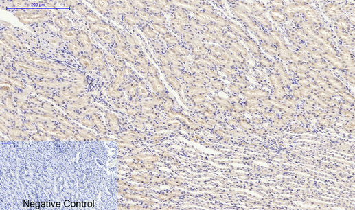
- Immunohistochemical analysis of paraffin-embedded Rat-kidney tissue. 1,HSP27 Polyclonal Antibody was diluted at 1:200(4°C,overnight). 2, Sodium citrate pH 6.0 was used for antibody retrieval(>98°C,20min). 3,Secondary antibody was diluted at 1:200(room tempeRature, 30min). Negative control was used by secondary antibody only.

- Western Blot analysis of 22RV1 cells using HSP27 Polyclonal Antibody diluted at 1:1000

- Immunohistochemistry analysis of paraffin-embedded human brain tissue, using HSP27 Antibody. The picture on the right is blocked with the synthesized peptide.
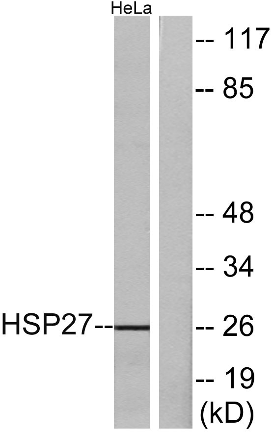
- Western blot analysis of lysates from HeLa cells, treated with Ca2+, using HSP27 Antibody. The lane on the right is blocked with the synthesized peptide.


