- 靶点:
- DRP1
- 简介:
- >>Necroptosis;>>NOD-like receptor signaling pathway;>>TNF signaling pathway
- 基因名称:
- DNM1L
- 蛋白名称:
- Dynamin-1-like protein
- Human Gene Id:
- 10059
- Human Swiss Prot No:
- O00429
- Mouse Gene Id:
- 74006
- Mouse Swiss Prot No:
- Q8K1M6
- Rat Gene Id:
- 114114
- Rat Swiss Prot No:
- O35303
- 免疫原:
- Synthesized peptide derived from DRP1 . at AA range: 580-660
- 特异性:
- DRP1 Polyclonal Antibody detects endogenous levels of DRP1 protein.
- 组成:
- Liquid in PBS containing 50% glycerol, 0.5% BSA and 0.02% sodium azide.
- 来源:
- Polyclonal, Rabbit,IgG
- 稀释:
- IF 1:50-200 WB 1:500 - 1:2000. IHC 1:100 - 1:300. ELISA: 1:10000. Not yet tested in other applications.
- 纯化工艺:
- The antibody was affinity-purified from rabbit antiserum by affinity-chromatography using epitope-specific immunogen.
- 浓度:
- 1 mg/ml
- 储存:
- -15°C to -25°C/1 year(Do not lower than -25°C)
- 其他名称:
- DNM1L;DLP1;DRP1;Dynamin-1-like protein;Dnm1p/Vps1p-like protein;DVLP;Dynamin family member proline-rich carboxyl-terminal domain less;Dymple;Dynamin-like protein;Dynamin-like protein 4;Dynamin-like protein IV;HdynIV;Dynamin-rela
- 实测条带:
- 80kD
- 背景:
- This gene encodes a member of the dynamin superfamily of GTPases. The encoded protein mediates mitochondrial and peroxisomal division, and is involved in developmentally regulated apoptosis and programmed necrosis. Dysfunction of this gene is implicated in several neurological disorders, including Alzheimer's disease. Mutations in this gene are associated with the autosomal dominant disorder, encephalopathy, lethal, due to defective mitochondrial and peroxisomal fission (EMPF). Alternative splicing results in multiple transcript variants encoding different isoforms. [provided by RefSeq, Jun 2013],
- 功能:
- catalytic activity:GTP + H(2)O = GDP + phosphate.,function:Functions in mitochondrial and peroxisomal division probably by regulating membrane fission. Enzyme hydrolyzing GTP that oligomerizes to form ring-like structures and is able to remodel membranes. May also play a role on organelles of the secretory pathway.,miscellaneous:Isoform 1 and isoform 2 inhibits peroxisomal division when overexpressed while isoform 3 and isoform 4 have no effect.,PTM:Phosphorylated by GSK3B.,similarity:Belongs to the dynamin family.,similarity:Contains 1 GED domain.,subcellular location:Mainly cytosolic. Also membrane-associated. Localizes to mitochondria at spots of division events. Associated with peroxisomal membranes, it is recruited in part by PEX11B. May also be associated with endoplasmic reticulum tubules and cytoplasmic vesicles and found to be perinuclear.,subunit:Homotetramer; N-terminal part b
- 细胞定位:
- Cytoplasm, cytosol. Golgi apparatus. Endomembrane system; Peripheral membrane protein. Mitochondrion outer membrane ; Peripheral membrane protein. Peroxisome. Membrane, clathrin-coated pit . Cytoplasmic vesicle, secretory vesicle, synaptic vesicle membrane . Mainly cytosolic. Recruited by RALA and RALBP1 to mitochondrion during mitosis (PubMed:21822277). Translocated to the mitochondrial membrane through O-GlcNAcylation and interaction with FIS1. Colocalized with MARCHF5 at mitochondrial membrane. Localizes to mitochondria at sites of division. Localizes to mitochondria following necrosis induction. Recruited to the mitochondrial outer membrane by interaction with MIEF1. Mitochondrial recruitment is inhibited by C11orf65/MFI (By similarity). Associated with peroxisomal membranes, partly re
- 组织表达:
- Ubiquitously expressed with highest levels found in skeletal muscles, heart, kidney and brain. Isoform 1 is brain-specific. Isoform 2 and isoform 3 are predominantly expressed in testis and skeletal muscles respectively. Isoform 4 is weakly expressed in brain, heart and kidney. Isoform 5 is dominantly expressed in liver, heart and kidney. Isoform 6 is expressed in neurons.
Mitochondrial separation protein inhibitor inhibits cell apoptosis in rat lungs during intermittent hypoxia. Experimental and Therapeutic Medicine 2019 Mar 01 WB Human,Rat 1:1000 lung WRTL1 cell
货号:YT1414
Experimental and therapeutic medicine 17.3 (2019): 2349-2358.
货号:YT1414
miR-34a/DRP-1-mediated mitophagy participated in cisplatin-induced ototoxicity via increasing oxidative stress BMC Pharmacology & Toxicology Haidi Yang WB Mouse cochlear tissue HEI-OC1 cell
货号:YT1414
PIM1 attenuates cisplatin-induced AKI by inhibiting Drp1 activation CELLULAR SIGNALLING Yuzhen Li WB Mouse 1:1000 kidney tissue mouse proximal tubular cell
货号:YT1414
ALCAT1-mediated abnormal cardiolipin remodelling promotes mitochondrial injury in podocytes in diabetic kidney disease Cell Communication and Signaling Hao Yiqun WB Human,Mouse 1:1000 glomerulus tissue podocytes
货号:YT1414
Human umbilical cord-derived mesenchymal stem cells ameliorate liver fibrosis by improving mitochondrial function via Slc25a47-Sirt3 signaling pathway BIOMEDICINE & PHARMACOTHERAPY Ping Chen IF Mouse liver tissue
货号:YT1414
- June 19-2018
- WESTERN IMMUNOBLOTTING PROTOCOL
- June 19-2018
- IMMUNOHISTOCHEMISTRY-PARAFFIN PROTOCOL
- June 19-2018
- IMMUNOFLUORESCENCE PROTOCOL
- September 08-2020
- FLOW-CYTOMEYRT-PROTOCOL
- May 20-2022
- Cell-Based ELISA│解您多样本WB检测之困扰
- July 13-2018
- CELL-BASED-ELISA-PROTOCOL-FOR-ACETYL-PROTEIN
- July 13-2018
- CELL-BASED-ELISA-PROTOCOL-FOR-PHOSPHO-PROTEIN
- July 13-2018
- Antibody-FAQs
- 产品图片
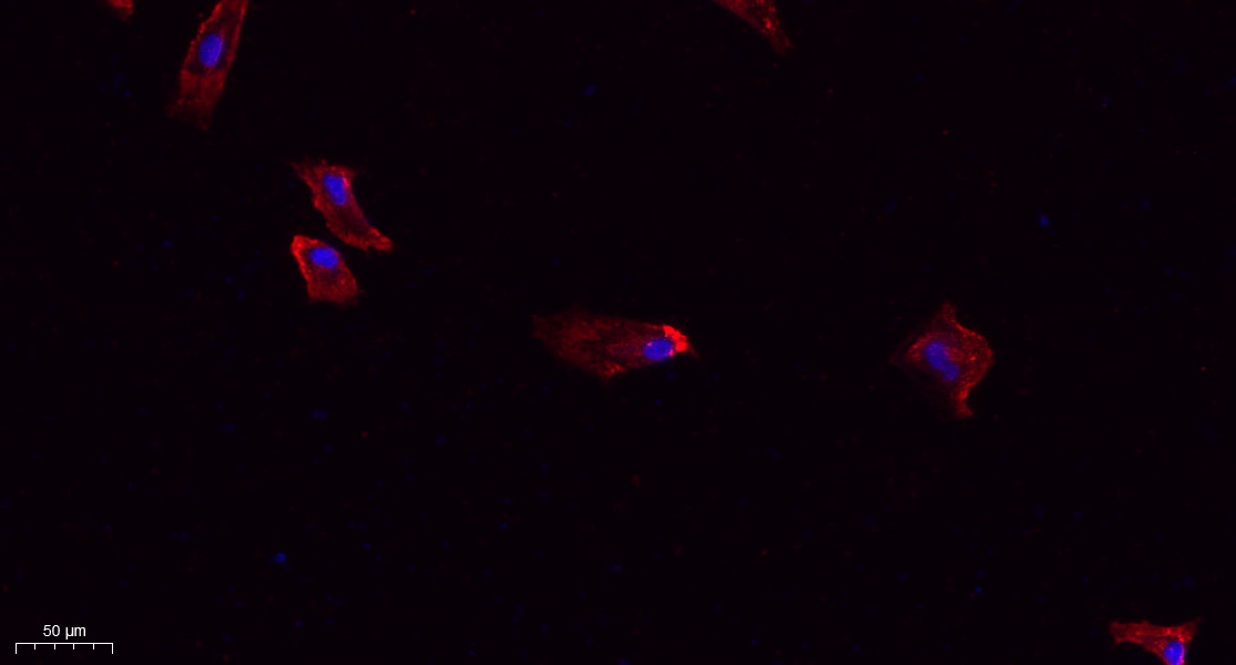
- Immunofluorescence analysis of A549. 1,primary Antibody(red) was diluted at 1:200(4°C overnight). 2, Goat Anti Rabbit IgG (H&L) - Alexa Fluor 594 Secondary antibody was diluted at 1:1000(room temperature, 50min).3, Picture B: DAPI(blue) 10min.

- Immunofluorescence analysis of Hela cell. 1,DRP1 Polyclonal Antibody(green) was diluted at 1:200(4° overnight). 2, Goat Anti Rabbit Alexa Fluor 488 Catalog:RS3211 was diluted at 1:1000(room temperature, 50min). 3 DAPI(blue) 10min.
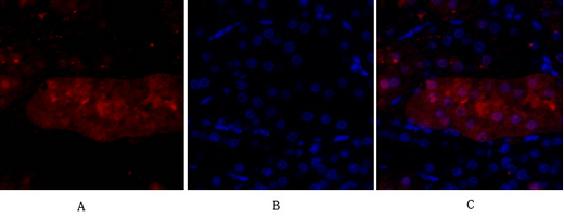
- Immunofluorescence analysis of rat-kidney tissue. 1,DRP1 Polyclonal Antibody(red) was diluted at 1:200(4°C,overnight). 2, Cy3 labled Secondary antibody was diluted at 1:300(room temperature, 50min).3, Picture B: DAPI(blue) 10min. Picture A:Target. Picture B: DAPI. Picture C: merge of A+B

- Immunohistochemical analysis of paraffin-embedded Rat-testis tissue. 1,DRP1 Polyclonal Antibody was diluted at 1:200(4°C,overnight). 2, Sodium citrate pH 6.0 was used for antibody retrieval(>98°C,20min). 3,Secondary antibody was diluted at 1:200(room tempeRature, 30min). Negative control was used by secondary antibody only.
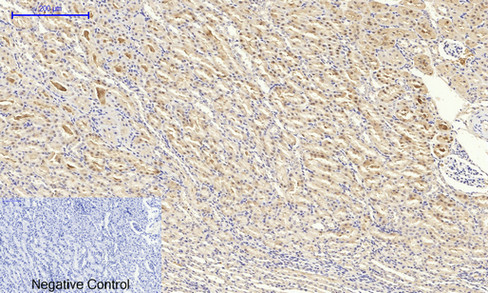
- Immunohistochemical analysis of paraffin-embedded Rat-kidney tissue. 1,DRP1 Polyclonal Antibody was diluted at 1:200(4°C,overnight). 2, Sodium citrate pH 6.0 was used for antibody retrieval(>98°C,20min). 3,Secondary antibody was diluted at 1:200(room tempeRature, 30min). Negative control was used by secondary antibody only.
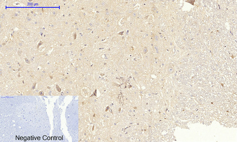
- Immunohistochemical analysis of paraffin-embedded Rat-spinal-cord tissue. 1,DRP1 Polyclonal Antibody was diluted at 1:200(4°C,overnight). 2, Sodium citrate pH 6.0 was used for antibody retrieval(>98°C,20min). 3,Secondary antibody was diluted at 1:200(room tempeRature, 30min). Negative control was used by secondary antibody only.
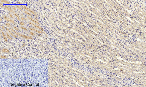
- Immunohistochemical analysis of paraffin-embedded Mouse-kidney tissue. 1,DRP1 Polyclonal Antibody was diluted at 1:200(4°C,overnight). 2, Sodium citrate pH 6.0 was used for antibody retrieval(>98°C,20min). 3,Secondary antibody was diluted at 1:200(room tempeRature, 30min). Negative control was used by secondary antibody only.

- Western Blot analysis of various cells using DRP1 Polyclonal Antibody diluted at 1:500
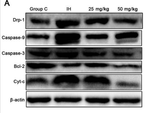
- Experimental and therapeutic medicine 17.3 (2019): 2349-2358.


