- 靶点:
- Cyclin B1
- 简介:
- >>FoxO signaling pathway;>>Cell cycle;>>Oocyte meiosis;>>p53 signaling pathway;>>Cellular senescence;>>Progesterone-mediated oocyte maturation;>>Human immunodeficiency virus 1 infection
- 基因名称:
- CCNB1
- 蛋白名称:
- G2/mitotic-specific cyclin-B1
- Human Gene Id:
- 891
- Human Swiss Prot No:
- P14635
- Mouse Gene Id:
- 268697
- Mouse Swiss Prot No:
- P24860
- Rat Gene Id:
- 25203
- Rat Swiss Prot No:
- P30277
- 免疫原:
- The antiserum was produced against synthesized peptide derived from human Cyclin B1. AA range:91-140
- 特异性:
- Cyclin B1 Polyclonal Antibody detects endogenous levels of Cyclin B1 protein.
- 组成:
- Liquid in PBS containing 50% glycerol, 0.5% BSA and 0.02% sodium azide.
- 来源:
- Polyclonal, Rabbit,IgG
- 稀释:
- WB 1:500 - 1:2000. IHC 1:100 - 1:300. IF 1:200 - 1:1000. ELISA: 1:20000. Not yet tested in other applications.
- 纯化工艺:
- The antibody was affinity-purified from rabbit antiserum by affinity-chromatography using epitope-specific immunogen.
- 浓度:
- 1 mg/ml
- 储存:
- -15°C to -25°C/1 year(Do not lower than -25°C)
- 其他名称:
- CCNB1;CCNB;G2/mitotic-specific cyclin-B1
- 实测条带:
- 60kD
- 背景:
- The protein encoded by this gene is a regulatory protein involved in mitosis. The gene product complexes with p34(cdc2) to form the maturation-promoting factor (MPF). Two alternative transcripts have been found, a constitutively expressed transcript and a cell cycle-regulated transcript, that is expressed predominantly during G2/M phase. The different transcripts result from the use of alternate transcription initiation sites. [provided by RefSeq, Jul 2008],
- 功能:
- developmental stage:Accumulates steadily during G2 and is abruptly destroyed at mitosis.,function:Essential for the control of the cell cycle at the G2/M (mitosis) transition.,PTM:Ubiquitinated by the SCF(NIPA) complex during interphase, leading to its destruction. Not ubiquitinated during G2/M phases.,similarity:Belongs to the cyclin family.,similarity:Belongs to the cyclin family. Cyclin AB subfamily.,subunit:Interacts with the CDC2 protein kinase to form a serine/threonine kinase holoenzyme complex also known as maturation promoting factor (MPF). The cyclin subunit imparts substrate specificity to the complex. Binds HEI10. Interacts with catalytically active RALBP1 and CDC2 during mitosis to form an endocytotic complex during interphase.,
- 细胞定位:
- Cytoplasm. Nucleus. Cytoplasm, cytoskeleton, microtubule organizing center, centrosome.
- 组织表达:
- Breast adenocarcinoma,Lung,Placenta,
Telekin suppresses human hepatocellular carcinoma cells in vitro by inducing G 2 /M phase arrest via the p38 MAPK signaling pathway. ACTA PHARMACOLOGICA SINICA Acta Pharmacol Sin. 2014 Sep;35(10):1311-1322 WB Human HepG2 cell
货号:YT1169
RhoB Acts as a Tumor Suppressor That Inhibits Malignancy of Clear Cell Renal Cell Carcinoma. PLoS One Plos One. 2016 Jul;11(7):e0157599 WB Human 1:1000 A498 cell, Caki-2 cell, 786-O cell, 769-P cell, Caki-1 cell
货号:YT1169
Li, Peng, et al. "Cell Cycle Arrest and Apoptosis Induction Activity of Nitidine Chloride on Acute Myeloid Leukemia Cells." Medicinal Chemistry 14.1 (2018): 60-66.
货号:YT1169
5, 6-Dihydroxy-3, 7, 4′-trimethoxyflavonol induces G2/M cell cycle arrest and apoptosis in human hepatocellular carcinoma cells." Journal of Asian natural products research 18.11 (2016): 1079-1090.
货号:YT1169
Nitidine chloride induces apoptosis, cell cycle arrest, and synergistic cytotoxicity with doxorubicin in breast cancer cells." Tumor Biology 35.10 (2014): 10201-10212.
货号:YT1169
Voacamine is a novel inhibitor of EGFR exerting oncogenic activity against colorectal cancer through the mitochondrial pathway PHARMACOLOGICAL RESEARCH Sanjun Shi WB Mouse
货号:YT1169
Protective effect of cornuside on OGD/R injury in SH-SY5Y cells and its underlying mechanism. BRAIN RESEARCH Jianwei Gong WB Human SH-SY5Y cell
货号:YT1169
- June 19-2018
- WESTERN IMMUNOBLOTTING PROTOCOL
- June 19-2018
- IMMUNOHISTOCHEMISTRY-PARAFFIN PROTOCOL
- June 19-2018
- IMMUNOFLUORESCENCE PROTOCOL
- September 08-2020
- FLOW-CYTOMEYRT-PROTOCOL
- May 20-2022
- Cell-Based ELISA│解您多样本WB检测之困扰
- July 13-2018
- CELL-BASED-ELISA-PROTOCOL-FOR-ACETYL-PROTEIN
- July 13-2018
- CELL-BASED-ELISA-PROTOCOL-FOR-PHOSPHO-PROTEIN
- July 13-2018
- Antibody-FAQs
- 产品图片

- Li, Lin, et al. "Telekin suppresses human hepatocellular carcinoma cells in vitro by inducing G 2/M phase arrest via the p38 MAPK signaling pathway." Acta Pharmacologica Sinica 35.10 (2014): 1311.
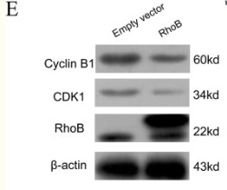
- Chen, Weihao, et al. "RhoB acts as a tumor suppressor that inhibits malignancy of clear cell renal cell carcinoma." PloS one 11.7 (2016): e0157599.

- Immunohistochemical analysis of paraffin-embedded Rat-heart tissue. 1,Cyclin B1 Polyclonal Antibody was diluted at 1:200(4°C,overnight). 2, Sodium citrate pH 6.0 was used for antibody retrieval(>98°C,20min). 3,Secondary antibody was diluted at 1:200(room tempeRature, 30min). Negative control was used by secondary antibody only.

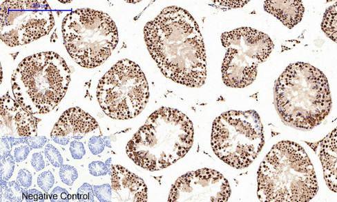
- Immunohistochemical analysis of paraffin-embedded Rat-testis tissue. 1,Cyclin B1 Polyclonal Antibody was diluted at 1:200(4°C,overnight). 2, Sodium citrate pH 6.0 was used for antibody retrieval(>98°C,20min). 3,Secondary antibody was diluted at 1:200(room tempeRature, 30min). Negative control was used by secondary antibody only.
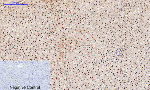
- Immunohistochemical analysis of paraffin-embedded Rat-liver tissue. 1,Cyclin B1 Polyclonal Antibody was diluted at 1:200(4°C,overnight). 2, Sodium citrate pH 6.0 was used for antibody retrieval(>98°C,20min). 3,Secondary antibody was diluted at 1:200(room tempeRature, 30min). Negative control was used by secondary antibody only.
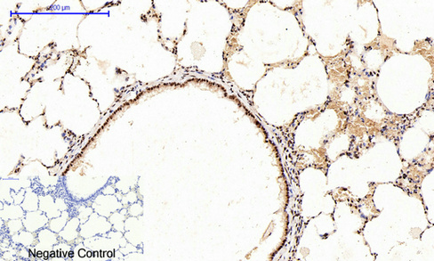
- Immunohistochemical analysis of paraffin-embedded Rat-lung tissue. 1,Cyclin B1 Polyclonal Antibody was diluted at 1:200(4°C,overnight). 2, Sodium citrate pH 6.0 was used for antibody retrieval(>98°C,20min). 3,Secondary antibody was diluted at 1:200(room tempeRature, 30min). Negative control was used by secondary antibody only.

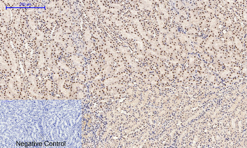
- Immunohistochemical analysis of paraffin-embedded Rat-kidney tissue. 1,Cyclin B1 Polyclonal Antibody was diluted at 1:200(4°C,overnight). 2, Sodium citrate pH 6.0 was used for antibody retrieval(>98°C,20min). 3,Secondary antibody was diluted at 1:200(room tempeRature, 30min). Negative control was used by secondary antibody only.
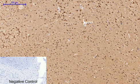
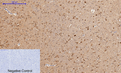
- Immunohistochemical analysis of paraffin-embedded Rat-brain tissue. 1,Cyclin B1 Polyclonal Antibody was diluted at 1:200(4°C,overnight). 2, Sodium citrate pH 6.0 was used for antibody retrieval(>98°C,20min). 3,Secondary antibody was diluted at 1:200(room tempeRature, 30min). Negative control was used by secondary antibody only.
- Immunohistochemical analysis of paraffin-embedded Mouse-testis tissue. 1,Cyclin B1 Polyclonal Antibody was diluted at 1:200(4°C,overnight). 2, Sodium citrate pH 6.0 was used for antibody retrieval(>98°C,20min). 3,Secondary antibody was diluted at 1:200(room tempeRature, 30min). Negative control was used by secondary antibody only.

- Immunohistochemical analysis of paraffin-embedded Mouse-liver tissue. 1,Cyclin B1 Polyclonal Antibody was diluted at 1:200(4°C,overnight). 2, Sodium citrate pH 6.0 was used for antibody retrieval(>98°C,20min). 3,Secondary antibody was diluted at 1:200(room tempeRature, 30min). Negative control was used by secondary antibody only.
- Immunohistochemical analysis of paraffin-embedded Mouse-kidney tissue. 1,Cyclin B1 Polyclonal Antibody was diluted at 1:200(4°C,overnight). 2, Sodium citrate pH 6.0 was used for antibody retrieval(>98°C,20min). 3,Secondary antibody was diluted at 1:200(room tempeRature, 30min). Negative control was used by secondary antibody only.
- Immunohistochemical analysis of paraffin-embedded Mouse-brain tissue. 1,Cyclin B1 Polyclonal Antibody was diluted at 1:200(4°C,overnight). 2, Sodium citrate pH 6.0 was used for antibody retrieval(>98°C,20min). 3,Secondary antibody was diluted at 1:200(room tempeRature, 30min). Negative control was used by secondary antibody only.
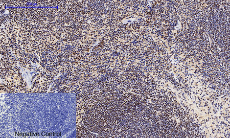
- Immunohistochemical analysis of paraffin-embedded Mouse-spleen tissue. 1,Cyclin B1 Polyclonal Antibody was diluted at 1:200(4°C,overnight). 2, Sodium citrate pH 6.0 was used for antibody retrieval(>98°C,20min). 3,Secondary antibody was diluted at 1:200(room tempeRature, 30min). Negative control was used by secondary antibody only.

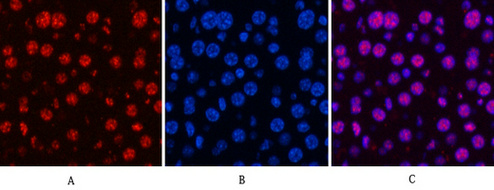
- Immunofluorescence analysis of Mouse-kidney tissue. 1,Cyclin B1 Polyclonal Antibody(red) was diluted at 1:200(4°C,overnight). 2, Cy3 labled Secondary antibody was diluted at 1:300(room temperature, 50min).3, Picture B: DAPI(blue) 10min. Picture A:Target. Picture B: DAPI. Picture C: merge of A+B

- Immunofluorescence analysis of Mouse-kidney tissue. 1,Cyclin B1 Polyclonal Antibody(red) was diluted at 1:200(4°C,overnight). 2, Cy3 labled Secondary antibody was diluted at 1:300(room temperature, 50min).3, Picture B: DAPI(blue) 10min. Picture A:Target. Picture B: DAPI. Picture C: merge of A+B
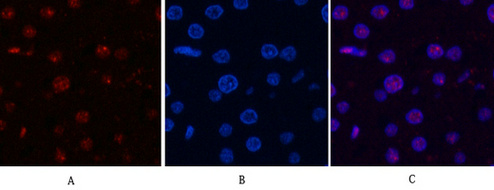
- Immunofluorescence analysis of Mouse-liver tissue. 1,Cyclin B1 Polyclonal Antibody(red) was diluted at 1:200(4°C,overnight). 2, Cy3 labled Secondary antibody was diluted at 1:300(room temperature, 50min).3, Picture B: DAPI(blue) 10min. Picture A:Target. Picture B: DAPI. Picture C: merge of A+B
- Immunofluorescence analysis of Rat-liver tissue. 1,Cyclin B1 Polyclonal Antibody(red) was diluted at 1:200(4°C,overnight). 2, Cy3 labled Secondary antibody was diluted at 1:300(room temperature, 50min).3, Picture B: DAPI(blue) 10min. Picture A:Target. Picture B: DAPI. Picture C: merge of A+B
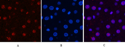
- Immunofluorescence analysis of Rat-liver tissue. 1,Cyclin B1 Polyclonal Antibody(red) was diluted at 1:200(4°C,overnight). 2, Cy3 labled Secondary antibody was diluted at 1:300(room temperature, 50min).3, Picture B: DAPI(blue) 10min. Picture A:Target. Picture B: DAPI. Picture C: merge of A+B
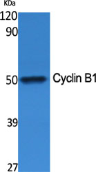
- Western Blot analysis of various cells using Cyclin B1 Polyclonal Antibody diluted at 1:500
.jpg)
- Western Blot analysis of PC-12 cells using Cyclin B1 Polyclonal Antibody diluted at 1:500

- Immunofluorescence analysis of HeLa cells, using Cyclin B1 Antibody. The picture on the right is blocked with the synthesized peptide.

- Immunohistochemistry analysis of paraffin-embedded human breast carcinoma tissue, using Cyclin B1 Antibody. The picture on the right is blocked with the synthesized peptide.
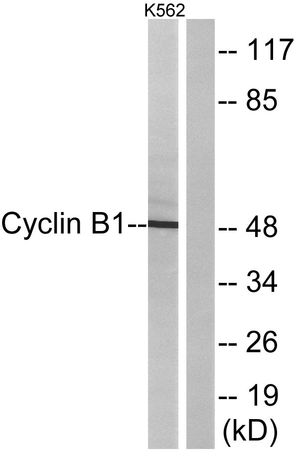
- Western blot analysis of lysates from K562 cells, treated with serum 10% 15', using Cyclin B1 Antibody. The lane on the right is blocked with the synthesized peptide.


