JNK1/2/3 (phospho Thr183) Polyclonal Antibody
- 货号:YP0156
- 应用:WB;IHC;IF;ELISA
- 种属:Human;Mouse;Rat;Chicken
- 靶点:
- JNK1/2/3
- 简介:
- >>Endocrine resistance;>>MAPK signaling pathway;>>ErbB signaling pathway;>>Ras signaling pathway;>>cAMP signaling pathway;>>FoxO signaling pathway;>>Sphingolipid signaling pathway;>>Mitophagy - animal;>>Autophagy - animal;>>Protein processing in endoplasmic reticulum;>>Apoptosis;>>Apoptosis - multiple species;>>Necroptosis;>>Wnt signaling pathway;>>Osteoclast differentiation;>>Focal adhesion;>>Tight junction;>>Toll-like receptor signaling pathway;>>NOD-like receptor signaling pathway;>>RIG-I-like receptor signaling pathway;>>C-type lectin receptor signaling pathway;>>IL-17 signaling pathway;>>Th1 and Th2 cell differentiation;>>Th17 cell differentiation;>>T cell receptor signaling pathway;>>Fc epsilon RI signaling pathway;>>TNF signaling pathway;>>Neurotrophin signaling pathway;>>Retrograde endocannabinoid signaling;>>Dopaminergic synapse;>>Inflammatory mediator regulation of TRP channels;>>Insulin signaling pathway;>>GnRH signaling pathway;>>Progesterone-mediated oocyte maturation;>>Pr
- 基因名称:
- MAPK8/9/10
- 蛋白名称:
- Mitogen-activated protein kinase 8/9/10
- Human Gene Id:
- 5599/5601/5602
- Human Swiss Prot No:
- P45983/P45984/P53779
- Mouse Gene Id:
- 26419/26420
- Rat Gene Id:
- 116554/50658/25272
- Rat Swiss Prot No:
- P49185/P49186/P49187
- 免疫原:
- The antiserum was produced against synthesized peptide derived from human SAPK/JNK around the phosphorylation site of Thr183. AA range:151-200
- 特异性:
- Phospho-JNK1/2/3 (T183) Polyclonal Antibody detects endogenous levels of JNK1/2/3 protein only when phosphorylated at T183.
- 组成:
- Liquid in PBS containing 50% glycerol, 0.5% BSA and 0.02% sodium azide.
- 来源:
- Polyclonal, Rabbit,IgG
- 稀释:
- WB 1:500 - 1:2000. IHC 1:100 - 1:300. IF 1:200 - 1:1000. ELISA: 1:5000. Not yet tested in other applications.
- 纯化工艺:
- The antibody was affinity-purified from rabbit antiserum by affinity-chromatography using epitope-specific immunogen.
- 浓度:
- 1 mg/ml
- 储存:
- -15°C to -25°C/1 year(Do not lower than -25°C)
- 其他名称:
- MAPK8;JNK1;PRKM8;SAPK1;SAPK1C;Mitogen-activated protein kinase 8;MAP kinase 8;MAPK 8;JNK-46;Stress-activated protein kinase 1c;SAPK1c;Stress-activated protein kinase JNK1;c-Jun N-terminal kinase 1;MAPK9;JNK2;PRKM9;SAPK1A;Mi
- 实测条带:
- 46kD,54kD
- 背景:
- The protein encoded by this gene is a member of the MAP kinase family. MAP kinases act as an integration point for multiple biochemical signals, and are involved in a wide variety of cellular processes such as proliferation, differentiation, transcription regulation and development. This kinase is activated by various cell stimuli, and targets specific transcription factors, and thus mediates immediate-early gene expression in response to cell stimuli. The activation of this kinase by tumor-necrosis factor alpha (TNF-alpha) is found to be required for TNF-alpha induced apoptosis. This kinase is also involved in UV radiation induced apoptosis, which is thought to be related to cytochrom c-mediated cell death pathway. Studies of the mouse counterpart of this gene suggested that this kinase play a key role in T cell proliferation, apoptosis and differentiation. Several alternatively spl
- 功能:
- catalytic activity:ATP + a protein = ADP + a phosphoprotein.,cofactor:Magnesium.,domain:The TXY motif contains the threonine and tyrosine residues whose phosphorylation activates the MAP kinases.,enzyme regulation:Activated by threonine and tyrosine phosphorylation by either of two dual specificity kinases, MAP2K4 and MAP2K7. Inhibited by dual specificity phosphatases, such as DUSP1.,function:JNK1 isoforms display different binding patterns: beta-1 preferentially binds to c-Jun, whereas alpha-1, alpha-2, and beta-2 have a similar low level of binding to both c-Jun or ATF2. However, there is no correlation between binding and phosphorylation, which is achieved at about the same efficiency by all isoforms.,function:Responds to activation by environmental stress and pro-inflammatory cytokines by phosphorylating a number of transcription factors, primarily components of AP-1 such as JUN, JDP
- 细胞定位:
- Cytoplasm . Nucleus . Cell junction, synapse . In the cortical neurons, predominantly cytoplasmic and associated with the Golgi apparatus and endosomal fraction. Increased neuronal activity increases phosphorylated form at synapses (By similarity). Colocalizes with POU5F1 in the nucleus. .
- 组织表达:
- Brain,Epithelium,Fetal brain,Lung,Pooled,Testis,
GCN5 participates in KLF4-VEGFA feedback to promote endometrial angiogenesis WB Human /HEMECs
货号:YP0156
Tamoxifen induces the development of hernia in mice by activating MMP-2 and MMP-13 expression. BIOCHIMICA ET BIOPHYSICA ACTA-MOLECULAR BASIS OF DISEASE 2015 Feb 19 WB Mouse NIH3T3 cell
货号:YP0156
The Effects of Maternal Atrazine Exposure and Swimming Training on Spatial Learning Memory and Hippocampal Morphology in Offspring Male Rats via PSD95/NR2B Signaling Pathway. CELLULAR AND MOLECULAR NEUROBIOLOGY Cell Mol Neurobiol. 2019 Oct;39(7):1003-1015 WB Rat 1:1000 hippocampus
货号:YP0156
The Role and Mechanism of SIRT1 in Resveratrol-regulated Osteoblast Autophagy in Osteoporosis Rats. Scientific Reports Sci Rep-Uk. 2019 Dec;9(1):1-15 WB Mouse osteoblasts
货号:YP0156
Inhibiting the Piezo1 channel protects microglia from acute hyperglycaemia damage through the JNK1 and mTOR signalling pathways. LIFE SCIENCES Life Sci. 2021 Jan;264:118667 WB Mouse BV2 cell
货号:YP0156
Telekin suppresses human hepatocellular carcinoma cells in vitro by inducing G 2 /M phase arrest via the p38 MAPK signaling pathway. ACTA PHARMACOLOGICA SINICA Acta Pharmacol Sin. 2014 Sep;35(10):1311-1322 WB Human HepG2 cell
货号:YP0156
Luo, Heguo, et al. "Yoda1 Activates Piezo1 in Vitro to Simulate the Upregulation of Piezo1 in the Infected Brain: Piezo1 Participates in the Immune Activation of Microglia." (2021).
货号:YP0156
Cadmium induces BNIP3-dependent autophagy in chicken spleen by modulating miR-33-AMPK axis. CHEMOSPHERE 2017 Dec 06 WB Chicken 1:1000 Spleen
货号:YP0156
Alpha-Momorcharin Inhibits Proinflammatory Cytokine Expression by M1 Macrophages but Not Anti-Inflammatory Cytokine Expression by M2 Macrophages Journal of Inflammation Research Fubing Shen WB Human
货号:YP0156
Morroniside Protects Human Granulosa Cells against H2O2-Induced Oxidative Damage by Regulating the Nrf2 and MAPK Signaling Pathways Evidence-based Complementary and Alternative Medicine Huilan Du WB Human
货号:YP0156
- June 19-2018
- WESTERN IMMUNOBLOTTING PROTOCOL
- June 19-2018
- IMMUNOHISTOCHEMISTRY-PARAFFIN PROTOCOL
- June 19-2018
- IMMUNOFLUORESCENCE PROTOCOL
- September 08-2020
- FLOW-CYTOMEYRT-PROTOCOL
- May 20-2022
- Cell-Based ELISA│解您多样本WB检测之困扰
- July 13-2018
- CELL-BASED-ELISA-PROTOCOL-FOR-ACETYL-PROTEIN
- July 13-2018
- CELL-BASED-ELISA-PROTOCOL-FOR-PHOSPHO-PROTEIN
- July 13-2018
- Antibody-FAQs
- 产品图片
.jpg)
- Jiao, Zhihui, et al. "Adipose-derived stem cells protect ischemia-reperfusion and partial hepatectomy by attenuating endoplasmic reticulum stress." Frontiers in cell and developmental biology 8 (2020): 177.
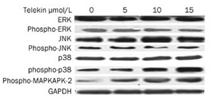
- Li, Lin, et al. "Telekin suppresses human hepatocellular carcinoma cells in vitro by inducing G 2/M phase arrest via the p38 MAPK signaling pathway." Acta Pharmacologica Sinica 35.10 (2014): 1311.
-if-62.jpg)
- Immunofluorescence analysis of rat-testis tissue. 1,JNK1/2/3 (phospho Thr183) Polyclonal Antibody(red) was diluted at 1:200(4°C,overnight). 2, Cy3 labled Secondary antibody was diluted at 1:300(room temperature, 50min).3, Picture B: DAPI(blue) 10min. Picture A:Target. Picture B: DAPI. Picture C: merge of A+B
-if-63.jpg)
- Immunofluorescence analysis of rat-testis tissue. 1,JNK1/2/3 (phospho Thr183) Polyclonal Antibody(red) was diluted at 1:200(4°C,overnight). 2, Cy3 labled Secondary antibody was diluted at 1:300(room temperature, 50min).3, Picture B: DAPI(blue) 10min. Picture A:Target. Picture B: DAPI. Picture C: merge of A+B
-if-64.jpg)
- Immunofluorescence analysis of rat-liver tissue. 1,JNK1/2/3 (phospho Thr183) Polyclonal Antibody(red) was diluted at 1:200(4°C,overnight). 2, Cy3 labled Secondary antibody was diluted at 1:300(room temperature, 50min).3, Picture B: DAPI(blue) 10min. Picture A:Target. Picture B: DAPI. Picture C: merge of A+B
-if-65.jpg)
- Immunofluorescence analysis of rat-liver tissue. 1,JNK1/2/3 (phospho Thr183) Polyclonal Antibody(red) was diluted at 1:200(4°C,overnight). 2, Cy3 labled Secondary antibody was diluted at 1:300(room temperature, 50min).3, Picture B: DAPI(blue) 10min. Picture A:Target. Picture B: DAPI. Picture C: merge of A+B
poly-ihc-rat-kidney.jpg)
- Immunohistochemical analysis of paraffin-embedded Rat-kidney tissue. 1,JNK1/2/3 (phospho Thr183) Polyclonal Antibody was diluted at 1:200(4°C,overnight). 2, Sodium citrate pH 6.0 was used for antibody retrieval(>98°C,20min). 3,Secondary antibody was diluted at 1:200(room tempeRature, 30min). Negative control was used by secondary antibody only.
poly-ihc-human-uterus.jpg)
- Immunohistochemical analysis of paraffin-embedded Human-uterus tissue. 1,JNK1/2/3 (phospho Thr183) Polyclonal Antibody was diluted at 1:200(4°C,overnight). 2, Sodium citrate pH 6.0 was used for antibody retrieval(>98°C,20min). 3,Secondary antibody was diluted at 1:200(room tempeRature, 30min). Negative control was used by secondary antibody only.
poly-ihc-human-uterus-cancer.jpg)
- Immunohistochemical analysis of paraffin-embedded Human-uterus-cancer tissue. 1,JNK1/2/3 (phospho Thr183) Polyclonal Antibody was diluted at 1:200(4°C,overnight). 2, Sodium citrate pH 6.0 was used for antibody retrieval(>98°C,20min). 3,Secondary antibody was diluted at 1:200(room tempeRature, 30min). Negative control was used by secondary antibody only.
poly-ihc-human-colon.jpg)
- Immunohistochemical analysis of paraffin-embedded Human-colon tissue. 1,JNK1/2/3 (phospho Thr183) Polyclonal Antibody was diluted at 1:200(4°C,overnight). 2, Sodium citrate pH 6.0 was used for antibody retrieval(>98°C,20min). 3,Secondary antibody was diluted at 1:200(room tempeRature, 30min). Negative control was used by secondary antibody only.
poly-ihc-human-liver.jpg)
- Immunohistochemical analysis of paraffin-embedded Human-liver tissue. 1,JNK1/2/3 (phospho Thr183) Polyclonal Antibody was diluted at 1:200(4°C,overnight). 2, Sodium citrate pH 6.0 was used for antibody retrieval(>98°C,20min). 3,Secondary antibody was diluted at 1:200(room tempeRature, 30min). Negative control was used by secondary antibody only.
poly-ihc-rat-spleen.jpg)
- Immunohistochemical analysis of paraffin-embedded Rat-spleen tissue. 1,JNK1/2/3 (phospho Thr183) Polyclonal Antibody was diluted at 1:200(4°C,overnight). 2, Sodium citrate pH 6.0 was used for antibody retrieval(>98°C,20min). 3,Secondary antibody was diluted at 1:200(room tempeRature, 30min). Negative control was used by secondary antibody only.
poly-ihc-mouse-heart.jpg)
- Immunohistochemical analysis of paraffin-embedded Mouse-heart tissue. 1,JNK1/2/3 (phospho Thr183) Polyclonal Antibody was diluted at 1:200(4°C,overnight). 2, Sodium citrate pH 6.0 was used for antibody retrieval(>98°C,20min). 3,Secondary antibody was diluted at 1:200(room tempeRature, 30min). Negative control was used by secondary antibody only.
poly-ihc-mouse-testis.jpg)
- Immunohistochemical analysis of paraffin-embedded Mouse-testis tissue. 1,JNK1/2/3 (phospho Thr183) Polyclonal Antibody was diluted at 1:200(4°C,overnight). 2, Sodium citrate pH 6.0 was used for antibody retrieval(>98°C,20min). 3,Secondary antibody was diluted at 1:200(room tempeRature, 30min). Negative control was used by secondary antibody only.
poly-ihc-mouse-colon.jpg)
- Immunohistochemical analysis of paraffin-embedded Mouse-colon tissue. 1,JNK1/2/3 (phospho Thr183) Polyclonal Antibody was diluted at 1:200(4°C,overnight). 2, Sodium citrate pH 6.0 was used for antibody retrieval(>98°C,20min). 3,Secondary antibody was diluted at 1:200(room tempeRature, 30min). Negative control was used by secondary antibody only.
poly-ihc-mouse-kidney.jpg)
- Immunohistochemical analysis of paraffin-embedded Mouse-kidney tissue. 1,JNK1/2/3 (phospho Thr183) Polyclonal Antibody was diluted at 1:200(4°C,overnight). 2, Sodium citrate pH 6.0 was used for antibody retrieval(>98°C,20min). 3,Secondary antibody was diluted at 1:200(room tempeRature, 30min). Negative control was used by secondary antibody only.
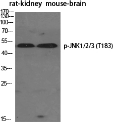
- Western Blot analysis of various cells using Phospho-JNK1/2/3 (T183) Polyclonal Antibody diluted at 1:1000
.jpg)
- Western Blot analysis of MCF-7 cells using Phospho-JNK1/2/3 (T183) Polyclonal Antibody diluted at 1:1000
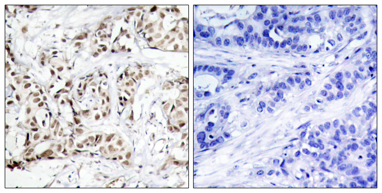
- Immunohistochemistry analysis of paraffin-embedded human breast carcinoma, using SAPK/JNK (Phospho-Thr183) Antibody. The picture on the right is blocked with the phospho peptide.
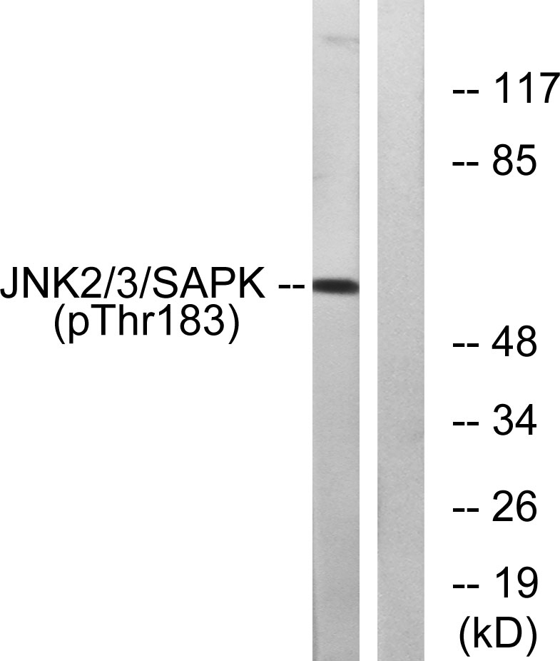
- Western blot analysis of lysates from HeLa cells treated with Anisomycin 200ng/ml 10', using SAPK/JNK (Phospho-Thr183) Antibody. The lane on the right is blocked with the phospho peptide.


