E-cadherin Monoclonal Antibody
- 货号:YM0207
- 应用:WB;IHC;IF;FCM;ELISA
- 种属:Human;Mouse;Monkey
- 靶点:
- E-cadherin
- 简介:
- >>Rap1 signaling pathway;>>Apelin signaling pathway;>>Hippo signaling pathway;>>Cell adhesion molecules;>>Adherens junction;>>Bacterial invasion of epithelial cells;>>Pathways in cancer;>>Endometrial cancer;>>Thyroid cancer;>>Melanoma;>>Bladder cancer;>>Gastric cancer
- 基因名称:
- CDH1
- 蛋白名称:
- Cadherin-1
- Human Gene Id:
- 999
- Human Swiss Prot No:
- P12830
- Mouse Gene Id:
- 12550
- Mouse Swiss Prot No:
- P09803
- 免疫原:
- Purified recombinant fragment of human E-cadherin expressed in E. Coli.
- 特异性:
- E-cadherin Monoclonal Antibody detects endogenous levels of E-cadherin protein.
- 组成:
- Liquid in PBS containing 50% glycerol, 0.5% BSA and 0.02% sodium azide.
- 来源:
- Monoclonal, Mouse
- 稀释:
- WB 1:500 - 1:2000. IHC 1:200 - 1:1000. Flow cytometry: 1:200 - 1:400. ELISA: 1:10000.. IF 1:50-200
- 纯化工艺:
- Affinity purification
- 储存:
- -15°C to -25°C/1 year(Do not lower than -25°C)
- 其他名称:
- CDH1;CDHE;UVO;Cadherin-1;CAM 120/80;Epithelial cadherin;E-cadherin;Uvomorulin;CD antigen CD324
- 实测条带:
- 125-130kD
- 背景:
- This gene encodes a classical cadherin of the cadherin superfamily. Alternative splicing results in multiple transcript variants, at least one of which encodes a preproprotein that is proteolytically processed to generate the mature glycoprotein. This calcium-dependent cell-cell adhesion protein is comprised of five extracellular cadherin repeats, a transmembrane region and a highly conserved cytoplasmic tail. Mutations in this gene are correlated with gastric, breast, colorectal, thyroid and ovarian cancer. Loss of function of this gene is thought to contribute to cancer progression by increasing proliferation, invasion, and/or metastasis. The ectodomain of this protein mediates bacterial adhesion to mammalian cells and the cytoplasmic domain is required for internalization. This gene is present in a gene cluster with other members of the cadherin family on chromosome 16. [provided by RefSeq, Nov 2015],
- 功能:
- disease:Defects in CDH1 are a cause of gastric cancer [MIM:137215]; also known as hereditary familial diffuse gastric cancer (HDGC).,disease:Defects in CDH1 are a cause of susceptibility to endometrial cancer [MIM:608089].,disease:Defects in CDH1 are associated with ovarian cancer [MIM:167000]. Ovarian cancer is the leading cause of death from gynecologic malignancy. It is characterized by advanced presentation with loco-regional dissemination in the peritoneal cavity and the rare incidence of visceral metastases. These typical features relate to the biology of the disease, which is a principal determinant of outcome.,disease:Defects in CDH1 are involved in dysfunction of the cell-cell adhesion system, triggering cancer invasion (gastric, breast, ovary, endometrium and thyroid) and metastasis.,function:Cadherins are calcium dependent cell adhesion proteins.,function:Cadherins are calcium
- 细胞定位:
- Cell junction, adherens junction . Cell membrane ; Single-pass type I membrane protein. Endosome. Golgi apparatus, trans-Golgi network. Colocalizes with DLGAP5 at sites of cell-cell contact in intestinal epithelial cells. Anchored to actin microfilaments through association with alpha-, beta- and gamma-catenin. Sequential proteolysis induced by apoptosis or calcium influx, results in translocation from sites of cell-cell contact to the cytoplasm. Colocalizes with RAB11A endosomes during its transport from the Golgi apparatus to the plasma membrane.
- 组织表达:
- Non-neural epithelial tissues.
The mechanism of mulberry leaves against renal tubular interstitial fibrosis through ERK1/2 signaling pathway was predicted by network pharmacology and validated in human tubular epithelial cells. PHYTOTHERAPY RESEARCH 2019 Jun 17 WB Human 1:800 HK-2 cell
货号:YM0207
- June 19-2018
- WESTERN IMMUNOBLOTTING PROTOCOL
- June 19-2018
- IMMUNOHISTOCHEMISTRY-PARAFFIN PROTOCOL
- June 19-2018
- IMMUNOFLUORESCENCE PROTOCOL
- September 08-2020
- FLOW-CYTOMEYRT-PROTOCOL
- May 20-2022
- Cell-Based ELISA│解您多样本WB检测之困扰
- July 13-2018
- CELL-BASED-ELISA-PROTOCOL-FOR-ACETYL-PROTEIN
- July 13-2018
- CELL-BASED-ELISA-PROTOCOL-FOR-PHOSPHO-PROTEIN
- July 13-2018
- Antibody-FAQs
- 产品图片
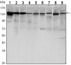
- Western Blot analysis using E-cadherin Monoclonal Antibody against LNCAP (1),A431 (2), DU145 (3), PC-3 (4), MCF-7 (5), PC-12 (6), NIH/3T3 (7), C6 (8) and COS7 (9) cell lysate.
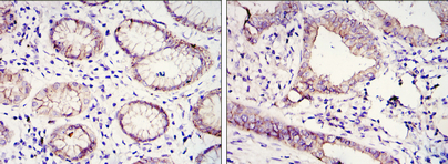
- Immunohistochemistry analysis of paraffin-embedded gastric cancer tissues (left) and lung cancer tissues (right) with DAB staining using E-cadherin Monoclonal Antibody.
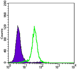
- Flow cytometric analysis of Hela cells using E-cadherin Monoclonal Antibody (green) and negative control (purple).


