LIMK-1/2 (PTR2545) Mouse mAb
- 货号:YM4686
- 应用:WB;ELISA
- 种属:Human; Mouse (predicted: Rat)
- 简介:
- >>Axon guidance;>>Fc gamma R-mediated phagocytosis;>>Regulation of actin cytoskeleton;>>Yersinia infection;>>Human immunodeficiency virus 1 infection
- 蛋白名称:
- LIM domain kinase 1 (LIMK-1) (EC 2.7.11.1)
- 免疫原:
- Synthesized peptide derived from human LIMK-1/2 AA range: 500-600
- 特异性:
- This antibody detects endogenous levels of LIMK-1/2 at Human, Mouse,Rat
- 组成:
- PBS, pH7.4, 50% glycerol, 0.03%Proclin 300
- 来源:
- Mouse,monoclonal:IgG1,Lambda
- 稀释:
- WB 1:500-2000 ELISA 1:5000-20000
- 储存:
- -15°C to -25°C/1 year(Do not lower than -25°C)
- 其他名称:
- LIM domain kinase 1 (LIMK-1) (EC 2.7.11.1)
- 背景:
- LIM domain kinase 1(LIMK1) Homo sapiens There are approximately 40 known eukaryotic LIM proteins, so named for the LIM domains they contain. LIM domains are highly conserved cysteine-rich structures containing 2 zinc fingers. Although zinc fingers usually function by binding to DNA or RNA, the LIM motif probably mediates protein-protein interactions. LIM kinase-1 and LIM kinase-2 belong to a small subfamily with a unique combination of 2 N-terminal LIM motifs and a C-terminal protein kinase domain. LIMK1 is a serine/threonine kinase that regulates actin polymerization via phosphorylation and inactivation of the actin binding factor cofilin. This protein is ubiquitously expressed during development and plays a role in many cellular processes associated with cytoskeletal structure. This protein also stimulates axon growth and may play a role in brain development. LIMK1 hemizygosity is implicated in the impaired visuospatial constructive cog
- 功能:
- catalytic activity:ATP + a protein = ADP + a phosphoprotein.,disease:Haploinsufficiency of LIMK1 may be the cause of certain cardiovascular and musculo-skeletal abnormalities observed in Williams-Beuren syndrome (WBS), a rare developmental disorder. It is a contiguous gene deletion syndrome involving genes from chromosome band 7q11.23.,function:Protein kinase which regulates actin filament dynamics. Phosphorylates and inactivates the actin binding/depolymerizing factor cofilin, thereby stabilizing the actin cytoskeleton. Isoform 3 has a dominant negative effect on actin cytoskeletal changes. May be involved in brain development.,PTM:Autophosphorylated.,PTM:Phosphorylated on serine and/or threonine residues by ROCK1. May be dephosphorylated and inactivated by SSH1.,similarity:Belongs to the protein kinase superfamily. TKL Ser/Thr protein kinase family.,similarity:Contains 1 PDZ (DHR) doma
- 细胞定位:
- Cytoplasm . Nucleus . Cytoplasm, cytoskeleton . Cell projection, lamellipodium . Predominantly found in the cytoplasm. Localizes in the lamellipodium in a CDC42BPA, CDC42BPB and FAM89B/LRAP25-dependent manner. .
- 组织表达:
- Highest expression in both adult and fetal nervous system. Detected ubiquitously throughout the different regions of adult brain, with highest levels in the cerebral cortex. Expressed to a lesser extent in heart and skeletal muscle.
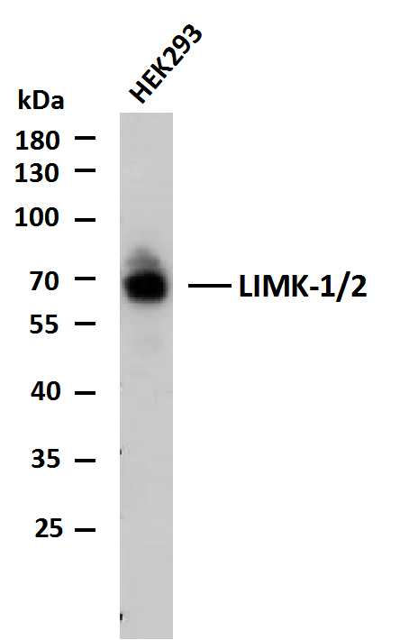
- Whole cell lysates of HEK293 were separated by 10% SDS-PAGE, and the membrane was blotted with anti-LIMK-1/2(PTR2545) antibody. The HRP-conjugated Goat anti-Mouse IgG(H + L) antibody was used to detect the antibody.
Lane 1: HEK293
Predicted band size: 65,72kDa
Observed band size: 68,72kDa
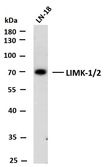
- Whole cell lysates of LN-18 were separated by 10% SDS-PAGE, and the membrane was blotted with anti-LIMK-1/2(PTR2545) antibody. The HRP-conjugated Goat anti-Mouse IgG(H + L) antibody was used to detect the antibody.
Lane 1: LN-18
Predicted band size: 65,72kDa
Observed band size: 70kDa
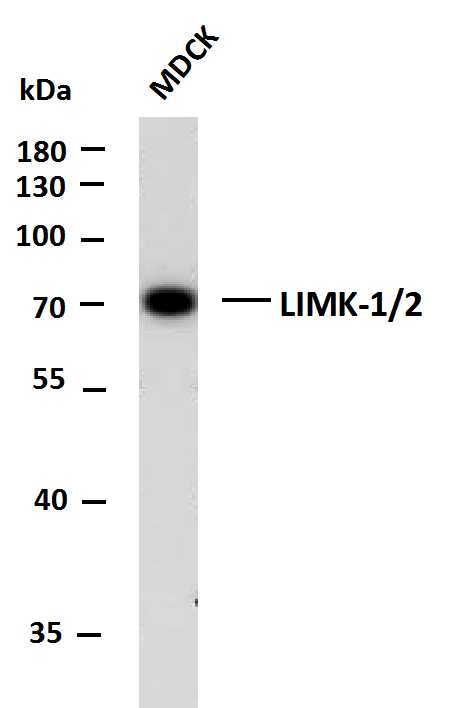
- Whole cell lysates of MDCK were separated by 10% SDS-PAGE, and the membrane was blotted with anti-LIMK-1/2(PTR2545) antibody. The HRP-conjugated Goat anti-Mouse IgG(H + L) antibody was used to detect the antibody.
Lane 1: MDCK
Predicted band size: 65,72kDa
Observed band size: 70kDa
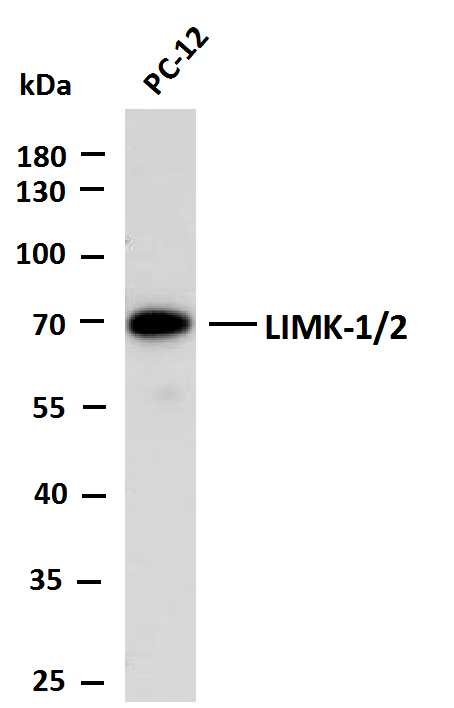
- Whole cell lysates of PC-12 were separated by 10% SDS-PAGE, and the membrane was blotted with anti-LIMK-1/2(PTR2545) antibody. The HRP-conjugated Goat anti-Mouse IgG(H + L) antibody was used to detect the antibody.
Lane 1: PC-12
Predicted band size: 65,72kDa
Observed band size: 70kDa
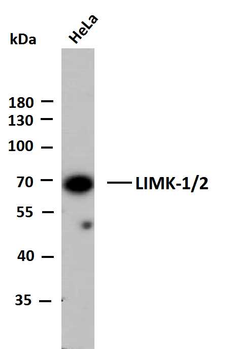
- Whole cell lysates of HeLa were separated by 10% SDS-PAGE, and the membrane was blotted with anti-LIMK-1/2 antibody. The HRP-conjugated Goat anti-Mouse IgG(H + L) antibody was used to detect the antibody.
Lane 1: HeLa
Predicted band size: 65,72kDa
Observed band size: 68kDa
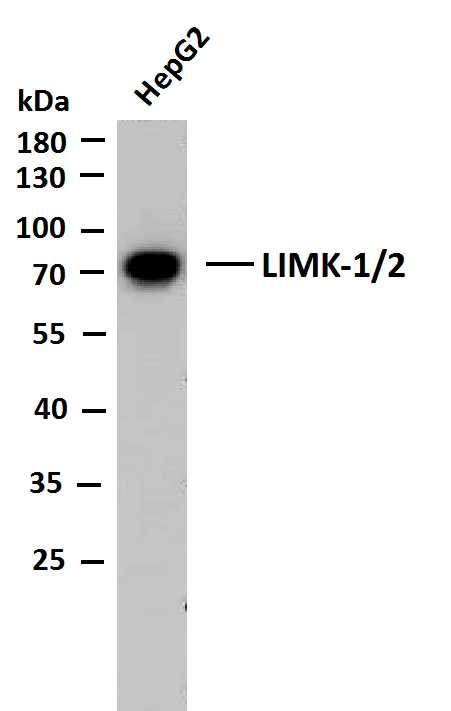
- Whole cell lysates of HepG2 were separated by 10% SDS-PAGE, and the membrane was blotted with anti-LIMK-1/2 antibody. The HRP-conjugated Goat anti-Mouse IgG(H + L) antibody was used to detect the antibody.
Lane 1: HepG2
Predicted band size: 65,72kDa
Observed band size: 72kDa
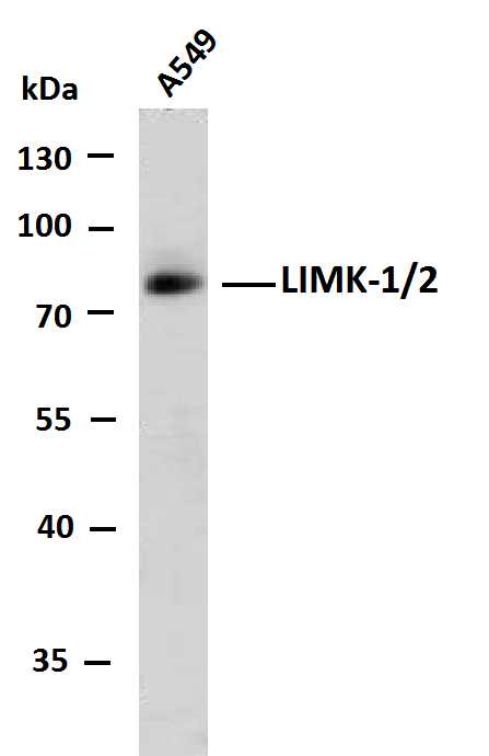
- Whole cell lysates of A549 were separated by 10% SDS-PAGE, and the membrane was blotted with anti-LIMK-1/2 antibody. The HRP-conjugated Goat anti-Mouse IgG(H + L) antibody was used to detect the antibody.
Lane 1: A549
Predicted band size: 65,72kDa
Observed band size: 72kDa


