


Catalog: YP1045
Size
Price
Status
Qty.
200μL
$600.00
In stock
0
100μL
$340.00
In stock
0
50μL
$190.00
In stock
0
Add to cart


Collected
Collect
Main Information
Target
IRAK-1
Host Species
Rabbit
Reactivity
Human, Mouse, Rat
Applications
IHC, IF, ELISA
MW
77kD (Calculated)
Conjugate/Modification
Phospho
Detailed Information
Recommended Dilution Ratio
IHC 1:100-1:300; ELISA 1:5000; IF 1:50-200
Formulation
Liquid in PBS containing 50% glycerol, 0.5% BSA and 0.02% sodium azide.
Specificity
Phospho-IRAK-1 (S376) Polyclonal Antibody detects endogenous levels of IRAK-1 protein only when phosphorylated at S376.The name of modified sites may be influenced by many factors, such as species (the modified site was not originally found in human samples) and the change of protein sequence (the previous protein sequence is incomplete, and the protein sequence may be prolonged with the development of protein sequencing technology). When naming, we will use the "numbers" in historical reference to keep the sites consistent with the reports. The antibody binds to the following modification sequence (lowercase letters are modification sites):QSsMV
Purification
The antibody was affinity-purified from rabbit antiserum by affinity-chromatography using epitope-specific immunogen.
Storage
-15°C to -25°C/1 year(Do not lower than -25°C)
Concentration
1 mg/ml
MW(Calculated)
77kD
Modification
Phospho
Clonality
Polyclonal
Isotype
IgG
Related Products
Secondary Antibodies
Goat Anti Mouse IgG(H+L) (HRP)
RS0001
More→
Secondary Antibodies

Goat Anti Rabbit IgG(H+L) (HRP)
RS0002
More→
Primary Antibodies
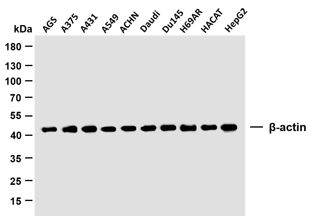
β-actin (PTR2364) Mouse mAb
YM3028
More→
Primary Antibodies
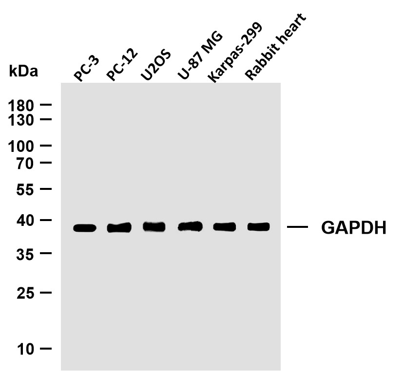
GAPDH (PTR2304) Mouse mAb
YM3029
More→
Antigen&Target Information
Immunogen:
The antiserum was produced against synthesized peptide derived from human IRAK1 around the phosphorylation site of Ser376. AA range:342-391
show all
Specificity:
Phospho-IRAK-1 (S376) Polyclonal Antibody detects endogenous levels of IRAK-1 protein only when phosphorylated at S376.The name of modified sites may be influenced by many factors, such as species (the modified site was not originally found in human samples) and the change of protein sequence (the previous protein sequence is incomplete, and the protein sequence may be prolonged with the development of protein sequencing technology). When naming, we will use the "numbers" in historical reference to keep the sites consistent with the reports. The antibody binds to the following modification sequence (lowercase letters are modification sites):QSsMV
show all
Gene Name:
IRAK1
show all
Protein Name:
Interleukin-1 receptor-associated kinase 1
show all
Other Name:
IRAK1 ;
IRAK ;
Interleukin-1 receptor-associated kinase 1 ;
IRAK-1
IRAK ;
Interleukin-1 receptor-associated kinase 1 ;
IRAK-1
show all
Background:
This gene encodes the interleukin-1 receptor-associated kinase 1, one of two putative serine/threonine kinases that become associated with the interleukin-1 receptor (IL1R) upon stimulation. This gene is partially responsible for IL1-induced upregulation of the transcription factor NF-kappa B. Alternatively spliced transcript variants encoding different isoforms have been found for this gene. [provided by RefSeq, Jul 2008],
show all
Function:
Catalytic activity:ATP + a protein = ADP + a phosphoprotein.,cofactor:Magnesium.,Function:Binds to the IL-1 type I receptor following IL-1 engagement, triggering intracellular signaling cascades leading to transcriptional up-regulation and mRNA stabilization. Isoform 1 binds rapidly but is then degraded allowing isoform 2 to mediate a slower, more sustained response to the cytokine. Isoform 2 is inactive suggesting that the kinase activity of this enzyme is not required for IL-1 signaling. Once phosphorylated, IRAK1 recruits the adapter protein PELI1.,PTM:Autophosphorylated or is transphosphorylated by IRAK4 following recruitment to the IL-1RI. In the case of isoform 1, this is linked to ubiquitination and degradation.,similarity:Belongs to the protein kinase superfamily.,similarity:Belongs to the protein kinase superfamily. TKL Ser/Thr protein kinase family. Pelle subfamily.,similarity:Contains 1 protein kinase domain.,subunit:IL-1 stimulation leads to the formation of a signaling complex which dissociates from the IL-1 receptor following the binding of PELI1. Interacts with IL1RL1. Interacts with IRAK1BP1.,tissue specificity:Isoform 1 and isoform 2 are ubiquitously expressed in all tissues examined, with isoform 1 being more strongly expressed than isoform 2.,
show all
Cellular Localization:
Cytoplasm . Nucleus . Lipid droplet . Translocates to the nucleus when sumoylated. RSAD2/viperin recruits it to the lipid droplet (By similarity). .
show all
Tissue Expression:
Research Areas:
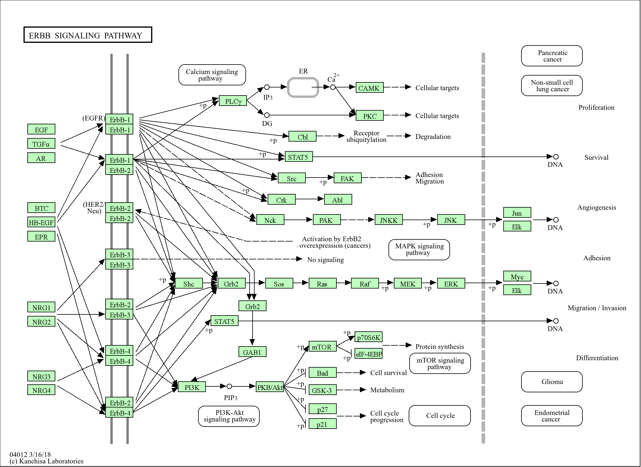
>>MAPK signaling pathway ;
>>NF-kappa B signaling pathway ;
>>Toll-like receptor signaling pathway ;
>>Neurotrophin signaling pathway ;
>>Alcoholic liver disease ;
>>Pathogenic Escherichia coli infection ;
>>Salmonella infection ;
>>Pertussis ;
>>Yersinia infection ;
>>Leishmaniasis ;
>>Chagas disease ;
>>Toxoplasmosis ;
>>Tuberculosis ;
>>Hepatitis B ;
>>Measles ;
>>Herpes simplex virus 1 infection ;
>>Epstein-Barr virus infection ;
>>Human immunodeficiency virus 1 infection ;
>>Coronavirus disease - COVID-19 ;
>>Lipid and atherosclerosis
show all
Signaling Pathway
Organismal Systems >> Immune system >> Toll-like receptor signaling pathway
Organismal Systems >> Nervous system >> Neurotrophin signaling pathway
Environmental Information Processing >> Signal transduction >> MAPK signaling pathway
Environmental Information Processing >> Signal transduction >> NF-kappa B signaling pathway
Reference Citation({{totalcount}})
Catalog: YP1045
Size
Price
Status
Qty.
200μL
$600.00
In stock
0
100μL
$340.00
In stock
0
50μL
$190.00
In stock
0
Add to cart
Collected
Collect
Recently Viewed Products
Clear allPRODUCTS
CUSTOMIZED
ABOUT US


Toggle night Mode
{{pinfoXq.title || ''}}
Catalog: {{pinfoXq.catalog || ''}}
Filter:
All
{{item.name}}
{{pinfo.title}}
-{{pinfo.catalog}}
Main Information
Target
{{pinfo.target}}
Reactivity
{{pinfo.react}}
Applications
{{pinfo.applicat}}
Conjugate/Modification
{{pinfo.coupling}}/{{pinfo.modific}}
MW (kDa)
{{pinfo.mwcalc}}
Host Species
{{pinfo.hostspec}}
Isotype
{{pinfo.isotype}}
Product {{index}}/{{pcount}}
Prev
Next
{{pvTitle}}
Scroll wheel zooms the picture
{{pvDescr}}