CYCS Polyclonal Antibody
- Catalog No.:YT5846
- Applications:IF;WB;IHC;ELISA
- Reactivity:Human;Mouse;Rat
- Target:
- Cytochrome C
- Fields:
- >>Oxidative phosphorylation;>>Metabolic pathways;>>Platinum drug resistance;>>p53 signaling pathway;>>Apoptosis;>>Apoptosis - multiple species;>>Non-alcoholic fatty liver disease;>>Alzheimer disease;>>Parkinson disease;>>Amyotrophic lateral sclerosis;>>Huntington disease;>>Spinocerebellar ataxia;>>Prion disease;>>Pathways of neurodegeneration - multiple diseases;>>Pathogenic Escherichia coli infection;>>Shigellosis;>>Salmonella infection;>>Legionellosis;>>Toxoplasmosis;>>Tuberculosis;>>Hepatitis C;>>Hepatitis B;>>Measles;>>Human cytomegalovirus infection;>>Influenza A;>>Kaposi sarcoma-associated herpesvirus infection;>>Herpes simplex virus 1 infection;>>Epstein-Barr virus infection;>>Human immunodeficiency virus 1 infection;>>Pathways in cancer;>>Colorectal cancer;>>Small cell lung cancer;>>Viral myocarditis;>>Lipid and atherosclerosis
- Gene Name:
- CYCS CYC
- Protein Name:
- CYCS
- Human Gene Id:
- 54205
- Human Swiss Prot No:
- P99999
- Mouse Gene Id:
- 13063
- Mouse Swiss Prot No:
- P62897
- Rat Swiss Prot No:
- P62898
- Immunogen:
- Synthetic peptide from human protein at AA range: 1-69
- Specificity:
- The antibody detects endogenous CYCS
- Formulation:
- Liquid in PBS containing 50% glycerol, 0.5% BSA and 0.02% sodium azide.
- Source:
- Polyclonal, Rabbit,IgG
- Dilution:
- IF 1:50-200 WB 1:500-2000,IHC 1:500-200, ELISA 1:10000-20000
- Purification:
- The antibody was affinity-purified from rabbit antiserum by affinity-chromatography using epitope-specific immunogen.
- Concentration:
- 1 mg/ml
- Storage Stability:
- -15°C to -25°C/1 year(Do not lower than -25°C)
- Other Name:
- Cytochrome c
- Observed Band(KD):
- 15kD
- Background:
- This gene encodes a small heme protein that functions as a central component of the electron transport chain in mitochondria. The encoded protein associates with the inner membrane of the mitochondrion where it accepts electrons from cytochrome b and transfers them to the cytochrome oxidase complex. This protein is also involved in initiation of apoptosis. Mutations in this gene are associated with autosomal dominant nonsyndromic thrombocytopenia. Numerous processed pseudogenes of this gene are found throughout the human genome.[provided by RefSeq, Jul 2010],
- Function:
- disease:Defects in CYCS are the cause of thrombocytopenia type 4 (THC4) [MIM:612004]; also known as autosomal dominant thrombocytopenia type 4. Thrombocytopenia is the presence of relatively few platelets in blood. THC4 is a non-syndromic form of thrombocytopenia. Clinical manifestations of thrombocytopenia are absent or mild. THC4 may be caused by dysregulated platelet formation.,function:Electron carrier protein. The oxidized form of the cytochrome c heme group can accept an electron from the heme group of the cytochrome c1 subunit of cytochrome reductase. Cytochrome c then transfers this electron to the cytochrome oxidase complex, the final protein carrier in the mitochondrial electron-transport chain.,function:Plays a role in apoptosis. Suppression of the anti-apoptotic members or activation of the pro-apoptotic members of the Bcl-2 family leads to altered mitochondrial membrane perm
- Subcellular Location:
- Mitochondrion intermembrane space. Loosely associated with the inner membrane.
- Expression:
- Amygdala,Bone marrow,Brain,Embryo,Heart,Kidney,Lung,Skeletal muscle,Skin,Testis,Uri
Licoflavone A Suppresses Gastric Cancer Growth and Metastasis by Blocking the VEGFR-2 Signaling Pathway. Journal of Oncology2022;2022:5497991. Human 1 : 1000 MKN-45 cell
The inhibitory effects of selenium nanoparticles modified by fructose-enriched polysaccharide from Codonopsis pilosula on HepG2 cells. INDUSTRIAL CROPS AND PRODUCTS Ind Crop Prod. 2022 Feb;176:114335 WB Human HepG2 cell
Curcumin Induces Apoptosis in SGC-7901 Gastric Adenocarcinoma Cells via Regulation of Mitochondrial Signaling Pathways. Asian Pacific Journal of Cancer Prevention 2014 May 15 WB Human SGC-7901 cel
Inhibition of mitochondrial VDAC1 oligomerization alleviates apoptosis and necroptosis of retinal neurons following OGD/R injury ANNALS OF ANATOMY-ANATOMISCHER ANZEIGER Wei-tao Yan WB Rat retinal tissues R28 retinal neuron-like cells
- June 19-2018
- WESTERN IMMUNOBLOTTING PROTOCOL
- June 19-2018
- IMMUNOHISTOCHEMISTRY-PARAFFIN PROTOCOL
- June 19-2018
- IMMUNOFLUORESCENCE PROTOCOL
- September 08-2020
- FLOW-CYTOMEYRT-PROTOCOL
- May 20-2022
- Cell-Based ELISA│解您多样本WB检测之困扰
- July 13-2018
- CELL-BASED-ELISA-PROTOCOL-FOR-ACETYL-PROTEIN
- July 13-2018
- CELL-BASED-ELISA-PROTOCOL-FOR-PHOSPHO-PROTEIN
- July 13-2018
- Antibody-FAQs
- Products Images

- Immunofluorescence analysis of mouse-spleen tissue. 1,CYCS Polyclonal Antibody(red) was diluted at 1:200(4°C,overnight). 2, Cy3 labled Secondary antibody was diluted at 1:300(room temperature, 50min).3, Picture B: DAPI(blue) 10min. Picture A:Target. Picture B: DAPI. Picture C: merge of A+B

- Immunofluorescence analysis of rat-lung tissue. 1,CYCS Polyclonal Antibody(red) was diluted at 1:200(4°C,overnight). 2, Cy3 labled Secondary antibody was diluted at 1:300(room temperature, 50min).3, Picture B: DAPI(blue) 10min. Picture A:Target. Picture B: DAPI. Picture C: merge of A+B
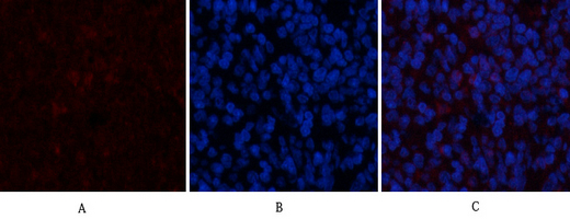
- Immunofluorescence analysis of rat-spleen tissue. 1,CYCS Polyclonal Antibody(red) was diluted at 1:200(4°C,overnight). 2, Cy3 labled Secondary antibody was diluted at 1:300(room temperature, 50min).3, Picture B: DAPI(blue) 10min. Picture A:Target. Picture B: DAPI. Picture C: merge of A+B

- Immunohistochemical analysis of paraffin-embedded Rat-kidney tissue. 1,CYCS Polyclonal Antibody was diluted at 1:200(4°C,overnight). 2, Sodium citrate pH 6.0 was used for antibody retrieval(>98°C,20min). 3,Secondary antibody was diluted at 1:200(room tempeRature, 30min). Negative control was used by secondary antibody only.
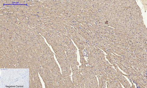
- Immunohistochemical analysis of paraffin-embedded Mouse-heart tissue. 1,CYCS Polyclonal Antibody was diluted at 1:200(4°C,overnight). 2, Sodium citrate pH 6.0 was used for antibody retrieval(>98°C,20min). 3,Secondary antibody was diluted at 1:200(room tempeRature, 30min). Negative control was used by secondary antibody only.
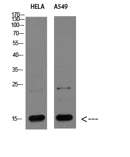
- Western blot analysis of HELA A549 Cell Lysate, antibody was diluted at 1:1000. Secondary antibody(catalog#:RS0002) was diluted at 1:20000

- Immunohistochemical analysis of paraffin-embedded human-muscle, antibody was diluted at 1:200


