XIAP (phospho Ser87) Polyclonal Antibody
- Catalog No.:YP0681
- Applications:WB;IHC;IF;ELISA
- Reactivity:Human;Mouse;Rat
- Target:
- XIAP
- Fields:
- >>Platinum drug resistance;>>NF-kappa B signaling pathway;>>Ubiquitin mediated proteolysis;>>Apoptosis;>>Apoptosis - multiple species;>>Necroptosis;>>Focal adhesion;>>NOD-like receptor signaling pathway;>>Toxoplasmosis;>>Human T-cell leukemia virus 1 infection;>>Pathways in cancer;>>Chemical carcinogenesis - receptor activation;>>Small cell lung cancer
- Gene Name:
- XIAP
- Protein Name:
- E3 ubiquitin-protein ligase XIAP
- Human Gene Id:
- 331
- Human Swiss Prot No:
- P98170
- Mouse Gene Id:
- 11798
- Mouse Swiss Prot No:
- Q60989
- Rat Gene Id:
- 63879
- Rat Swiss Prot No:
- Q9R0I6
- Immunogen:
- The antiserum was produced against synthesized peptide derived from human XIAP around the phosphorylation site of Ser87. AA range:53-102
- Specificity:
- Phospho-XIAP (S87) Polyclonal Antibody detects endogenous levels of XIAP protein only when phosphorylated at S87.
- Formulation:
- Liquid in PBS containing 50% glycerol, 0.5% BSA and 0.02% sodium azide.
- Source:
- Polyclonal, Rabbit,IgG
- Dilution:
- WB 1:500 - 1:2000. IHC 1:100 - 1:300. ELISA: 1:20000.. IF 1:50-200
- Purification:
- The antibody was affinity-purified from rabbit antiserum by affinity-chromatography using epitope-specific immunogen.
- Concentration:
- 1 mg/ml
- Storage Stability:
- -15°C to -25°C/1 year(Do not lower than -25°C)
- Other Name:
- XIAP;API3;BIRC4;IAP3;E3 ubiquitin-protein ligase XIAP;Baculoviral IAP repeat-containing protein 4;IAP-like protein;ILP;hILP;Inhibitor of apoptosis protein 3;IAP-3;hIAP-3;hIAP3;X-linked inhibitor of apoptosis protein;X-linked I
- Observed Band(KD):
- 57kD
- Background:
- This gene encodes a protein that belongs to a family of apoptotic suppressor proteins. Members of this family share a conserved motif termed, baculovirus IAP repeat, which is necessary for their anti-apoptotic function. This protein functions through binding to tumor necrosis factor receptor-associated factors TRAF1 and TRAF2 and inhibits apoptosis induced by menadione, a potent inducer of free radicals, and interleukin 1-beta converting enzyme. This protein also inhibits at least two members of the caspase family of cell-death proteases, caspase-3 and caspase-7. Mutations in this gene are the cause of X-linked lymphoproliferative syndrome. Alternate splicing results in multiple transcript variants. Pseudogenes of this gene are found on chromosomes 2 and 11.[provided by RefSeq, Feb 2011],
- Function:
- disease:Defects in XIAP are the cause of lymphoproliferative syndrome X-linked type 2 (XLP2) [MIM:300635]. XLP is a rare immunodeficiency characterized by extreme susceptibility to infection with Epstein-Barr virus (EBV). Symptoms include severe or fatal mononucleosis, acquired hypogammaglobulinemia, pancytopenia and malignant lymphoma.,domain:The first BIR domain is involved in interaction with MAP3K7IP1 and is important for dimerization. The second BIR domain is sufficient to inhibit caspase-3 and caspase-7, while the third BIR is involved in caspase-9 inhibition. The interactions with SMAC and PRSS25 are mediated by the second and third BIR domains.,function:Apoptotic suppressor. Has E3 ubiquitin-protein ligase activity. Mediates the proteasomal degradation of target proteins, such as caspase-3, SMAC or AIFM1. Inhibitor of caspase-3, -7 and -9. Mediates activation of MAP3K7/TAK1, lead
- Subcellular Location:
- Cytoplasm. Nucleus. TLE3 promotes its nuclear localization.
- Expression:
- Expressed in colonic crypts (at protein level) (PubMed:30389919). Ubiquitous, except peripheral blood leukocytes (PubMed:8654366).
Brucella Outer Membrane Lipoproteins 19 and 16 Differentially Induce Interleukin-18 Response or Pyroptosis in Human Monocytic Cells. JOURNAL OF INFECTIOUS DISEASES J Infect Dis. 2021 Dec;224(12):2148-2159 WB Human THP-1 cells
- June 19-2018
- WESTERN IMMUNOBLOTTING PROTOCOL
- June 19-2018
- IMMUNOHISTOCHEMISTRY-PARAFFIN PROTOCOL
- June 19-2018
- IMMUNOFLUORESCENCE PROTOCOL
- September 08-2020
- FLOW-CYTOMEYRT-PROTOCOL
- May 20-2022
- Cell-Based ELISA│解您多样本WB检测之困扰
- July 13-2018
- CELL-BASED-ELISA-PROTOCOL-FOR-ACETYL-PROTEIN
- July 13-2018
- CELL-BASED-ELISA-PROTOCOL-FOR-PHOSPHO-PROTEIN
- July 13-2018
- Antibody-FAQs
- Products Images
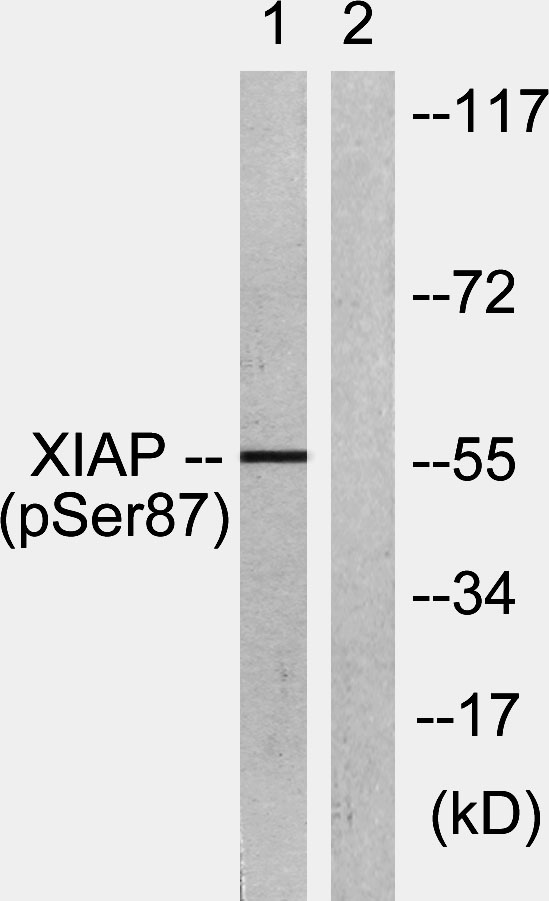
- Western Blot analysis of HepG2 cells using Phospho-XIAP (S87) Polyclonal Antibody diluted at 1:500
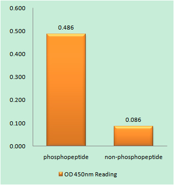
- Enzyme-Linked Immunosorbent Assay (Phospho-ELISA) for Immunogen Phosphopeptide (Phospho-left) and Non-Phosphopeptide (Phospho-right), using XIAP (Phospho-Ser87) Antibody
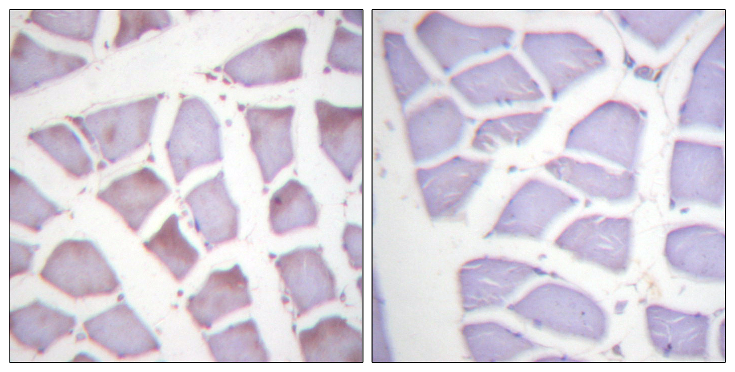
- Immunohistochemistry analysis of paraffin-embedded human skeletal muscle, using XIAP (Phospho-Ser87) Antibody. The picture on the right is blocked with the phospho peptide.
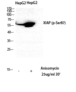
- Western blot analysis of lysates from HepG2 cells treated with Anisomycin 25ug/ml 30', using XIAP (Phospho-Ser87) Antibody. The lane on the right is blocked with the phospho peptide.