FADD (phospho Ser194) Polyclonal Antibody
- Catalog No.:YP0628
- Applications:WB;IHC;IF;ELISA
- Reactivity:Human;Mouse
- Target:
- FADD
- Fields:
- >>Platinum drug resistance;>>Apoptosis;>>Apoptosis - multiple species;>>Necroptosis;>>Toll-like receptor signaling pathway;>>NOD-like receptor signaling pathway;>>RIG-I-like receptor signaling pathway;>>IL-17 signaling pathway;>>TNF signaling pathway;>>Alcoholic liver disease;>>Alzheimer disease;>>Pathways of neurodegeneration - multiple diseases;>>Pathogenic Escherichia coli infection;>>Salmonella infection;>>Chagas disease;>>Tuberculosis;>>Hepatitis C;>>Hepatitis B;>>Measles;>>Human cytomegalovirus infection;>>Influenza A;>>Human papillomavirus infection;>>Kaposi sarcoma-associated herpesvirus infection;>>Herpes simplex virus 1 infection;>>Epstein-Barr virus infection;>>Human immunodeficiency virus 1 infection;>>Pathways in cancer
- Gene Name:
- FADD
- Protein Name:
- Protein FADD
- Human Gene Id:
- 8772
- Human Swiss Prot No:
- Q13158
- Mouse Swiss Prot No:
- Q61160
- Immunogen:
- The antiserum was produced against synthesized peptide derived from human FADD around the phosphorylation site of Ser194. AA range:159-208
- Specificity:
- Phospho-FADD (S194) Polyclonal Antibody detects endogenous levels of FADD protein only when phosphorylated at S194.
- Formulation:
- Liquid in PBS containing 50% glycerol, 0.5% BSA and 0.02% sodium azide.
- Source:
- Polyclonal, Rabbit,IgG
- Dilution:
- WB 1:500 - 1:2000. IHC 1:100 - 1:300. ELISA: 1:5000.. IF 1:50-200
- Purification:
- The antibody was affinity-purified from rabbit antiserum by affinity-chromatography using epitope-specific immunogen.
- Concentration:
- 1 mg/ml
- Storage Stability:
- -15°C to -25°C/1 year(Do not lower than -25°C)
- Other Name:
- FADD;MORT1;GIG3;Protein FADD;FAS-associated death domain protein;FAS-associating death domain-containing protein;Growth-inhibiting gene 3 protein;Mediator of receptor induced toxicity
- Observed Band(KD):
- 28kD
- Background:
- The protein encoded by this gene is an adaptor molecule that interacts with various cell surface receptors and mediates cell apoptotic signals. Through its C-terminal death domain, this protein can be recruited by TNFRSF6/Fas-receptor, tumor necrosis factor receptor, TNFRSF25, and TNFSF10/TRAIL-receptor, and thus it participates in the death signaling initiated by these receptors. Interaction of this protein with the receptors unmasks the N-terminal effector domain of this protein, which allows it to recruit caspase-8, and thereby activate the cysteine protease cascade. Knockout studies in mice also suggest the importance of this protein in early T cell development. [provided by RefSeq, Jul 2008],
- Function:
- domain:Contains a death domain involved in the binding of the corresponding domain within Fas receptor.,function:Apoptotic adaptor molecule that recruits caspase-8 or caspase-10 to the activated Fas (CD95) or TNFR-1 receptors. The resulting aggregate called the death-inducing signaling complex (DISC) performs caspase-8 proteolytic activation. Active caspase-8 initiates the subsequent cascade of caspases mediating apoptosis.,PTM:Phosphorylated.,similarity:Contains 1 death domain.,similarity:Contains 1 DED (death effector) domain.,subunit:Interacts with CFLAR, PEA15 and MBD4. When phosphorylated, part of a complex containing HIPK3 and FAS. May interact with MAVS/IPS1. Interacts with MOCV v-CFLAR protein and LRDD.,tissue specificity:Expressed in a wide variety of tissues, except for peripheral blood mononuclear leukocytes.,
- Subcellular Location:
- cytoplasm,cytosol,plasma membrane,death-inducing signaling complex,CD95 death-inducing signaling complex,neuron projection,cell body,membrane raft,ripoptosome,
- Expression:
- Expressed in a wide variety of tissues, except for peripheral blood mononuclear leukocytes.
- June 19-2018
- WESTERN IMMUNOBLOTTING PROTOCOL
- June 19-2018
- IMMUNOHISTOCHEMISTRY-PARAFFIN PROTOCOL
- June 19-2018
- IMMUNOFLUORESCENCE PROTOCOL
- September 08-2020
- FLOW-CYTOMEYRT-PROTOCOL
- May 20-2022
- Cell-Based ELISA│解您多样本WB检测之困扰
- July 13-2018
- CELL-BASED-ELISA-PROTOCOL-FOR-ACETYL-PROTEIN
- July 13-2018
- CELL-BASED-ELISA-PROTOCOL-FOR-PHOSPHO-PROTEIN
- July 13-2018
- Antibody-FAQs
- Products Images
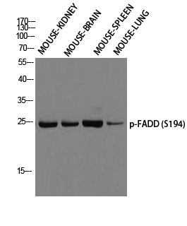
- Western blot analysis of MOUSE-KIDNEY MOUSE-BRAIN MOUSE-SPLEEN MOUSE-LUNG using p-FADD (S194) antibody. Antibody was diluted at 1:1000
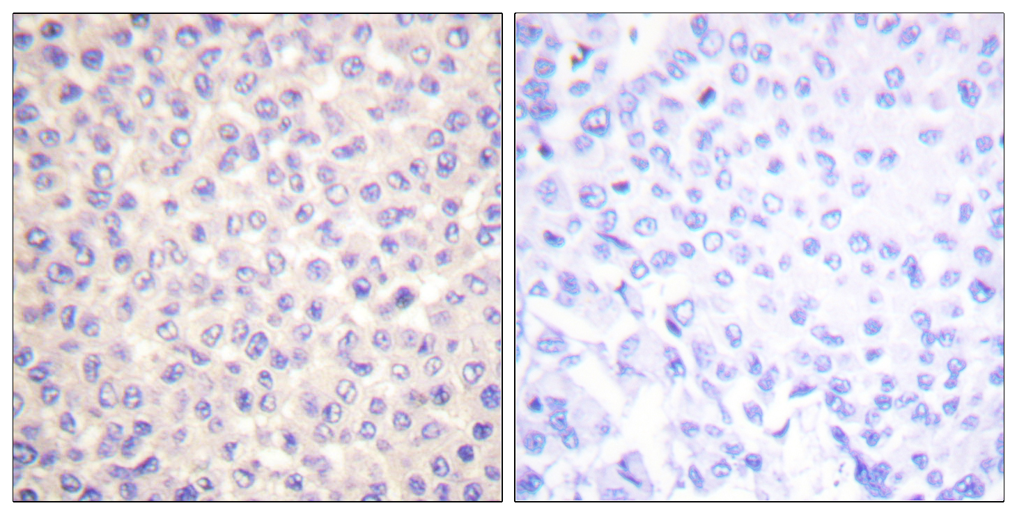
- Immunohistochemistry analysis of paraffin-embedded human breast carcinoma, using FADD (Phospho-Ser194) Antibody. The picture on the right is blocked with the phospho peptide.
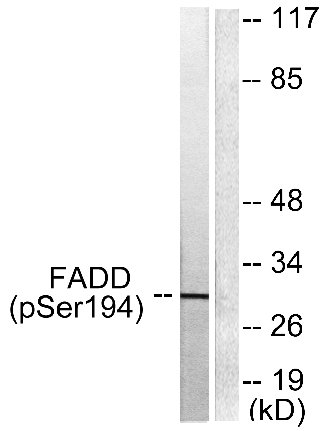
- Western blot analysis of lysates from HeLa cells treated with Paclitaxel 1uM 60', using FADD (Phospho-Ser194) Antibody. The lane on the right is blocked with the phospho peptide.


