PPAR-α Polyclonal Antibody
- Catalog No.:YT3835
- Applications:WB;IHC;IF;ELISA
- Reactivity:Human;Mouse;Rat
- Target:
- PPAR α
- Fields:
- >>PPAR signaling pathway;>>cAMP signaling pathway;>>Adipocytokine signaling pathway;>>Glucagon signaling pathway;>>Insulin resistance;>>Non-alcoholic fatty liver disease;>>Alcoholic liver disease;>>Hepatitis C;>>Chemical carcinogenesis - receptor activation;>>Diabetic cardiomyopathy
- Gene Name:
- PPARA
- Protein Name:
- Peroxisome proliferator-activated receptor alpha
- Human Gene Id:
- 5465
- Human Swiss Prot No:
- Q07869
- Mouse Gene Id:
- 19013
- Mouse Swiss Prot No:
- P23204
- Rat Gene Id:
- 25747
- Rat Swiss Prot No:
- P37230
- Immunogen:
- The antiserum was produced against synthesized peptide derived from human PPAR-alpha. AA range:6-55
- Specificity:
- PPAR-α Polyclonal Antibody detects endogenous levels of PPAR-α protein.
- Formulation:
- Liquid in PBS containing 50% glycerol, 0.5% BSA and 0.02% sodium azide.
- Source:
- Polyclonal, Rabbit,IgG
- Dilution:
- WB 1:500-2000, ELISA 1:10000-20000 IHC 1:50-300. IF 1:50-200
- Purification:
- The antibody was affinity-purified from rabbit antiserum by affinity-chromatography using epitope-specific immunogen.
- Concentration:
- 1 mg/ml
- Storage Stability:
- -15°C to -25°C/1 year(Do not lower than -25°C)
- Other Name:
- PPARA;NR1C1;PPAR;Peroxisome proliferator-activated receptor alpha;PPAR-alpha;Nuclear receptor subfamily 1 group C member 1
- Observed Band(KD):
- 52kD
- Background:
- peroxisome proliferator activated receptor alpha(PPARA) Homo sapiens Peroxisome proliferators include hypolipidemic drugs, herbicides, leukotriene antagonists, and plasticizers; this term arises because they induce an increase in the size and number of peroxisomes. Peroxisomes are subcellular organelles found in plants and animals that contain enzymes for respiration and for cholesterol and lipid metabolism. The action of peroxisome proliferators is thought to be mediated via specific receptors, called PPARs, which belong to the steroid hormone receptor superfamily. PPARs affect the expression of target genes involved in cell proliferation, cell differentiation and in immune and inflammation responses. Three closely related subtypes (alpha, beta/delta, and gamma) have been identified. This gene encodes the subtype PPAR-alpha, which is a nuclear transcription factor. Multiple alternatively spliced transcript variants have been described for thi
- Function:
- function:Receptor that binds peroxisome proliferators such as hypolipidemic drugs and fatty acids. Once activated by a ligand, the receptor binds to a promoter element in the gene for acyl-CoA oxidase and activates its transcription. It therefore controls the peroxisomal beta-oxidation pathway of fatty acids.,online information:Peroxisome proliferator-activated receptor entry,similarity:Belongs to the nuclear hormone receptor family. NR1 subfamily.,similarity:Contains 1 nuclear receptor DNA-binding domain.,subunit:Heterodimer with the retinoid X receptor. Interacts with NCOA3 and NCOA6 coactivators, leading to a strong increase of transcription of target genes. Also interacts with PPARBP coactivator in vitro. Interacts with AKAP13.,tissue specificity:Skeletal muscle, liver, heart and kidney.,
- Subcellular Location:
- Nucleus.
- Expression:
- Skeletal muscle, liver, heart and kidney. Expressed in monocytes (PubMed:28167758).
HtrA2/Omi mitigates NAFLD in high-fat-fed mice by ameliorating mitochondrial dysfunction and restoring autophagic flux. Cell Death Discovery2022 Apr;8(1):1-12. Mouse 1:100,1:1000 liver tissues
Oat Fiber Modulates Hepatic Circadian Clock via Promoting Gut Microbiota-Derived Short Chain Fatty Acids. JOURNAL OF AGRICULTURAL AND FOOD CHEMISTRY J Agr Food Chem. 2021;69(51):15624–15635 WB Mouse 1:1000 Liver
Rg1 Attenuates alcoholic hepatic damage through regulating AMP-activated protein kinase and nuclear factor erythroid 2-related factor 2 signal pathways. JOURNAL OF ASIAN NATURAL PRODUCTS RESEARCH 2016 May 26 WB Mouse liver
Apical papilla stem cell-derived exosomes regulate lipid metabolism and alleviate inflammation in the MCD-induced mouse NASH model BIOCHEMICAL PHARMACOLOGY Yi-Fei Nie WB Mouse 1:1000 liver tissue
- June 19-2018
- WESTERN IMMUNOBLOTTING PROTOCOL
- June 19-2018
- IMMUNOHISTOCHEMISTRY-PARAFFIN PROTOCOL
- June 19-2018
- IMMUNOFLUORESCENCE PROTOCOL
- September 08-2020
- FLOW-CYTOMEYRT-PROTOCOL
- May 20-2022
- Cell-Based ELISA│解您多样本WB检测之困扰
- July 13-2018
- CELL-BASED-ELISA-PROTOCOL-FOR-ACETYL-PROTEIN
- July 13-2018
- CELL-BASED-ELISA-PROTOCOL-FOR-PHOSPHO-PROTEIN
- July 13-2018
- Antibody-FAQs
- Products Images
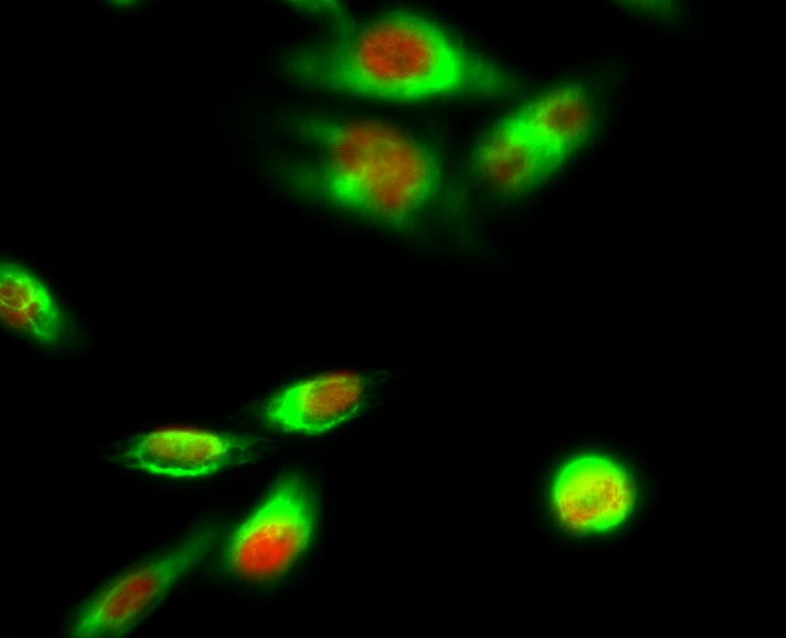
- Immunofluorescence analysis of Hela cell. 1,PPAR-α Polyclonal Antibody(red) was diluted at 1:200(4° overnight). Galectin-3 Monoclonal Antibody(6G2)(green) was diluted at 1:200(4° overnight). 2, Goat Anti Rabbit Alexa Fluor 594 Catalog:RS3611 was diluted at 1:1000(room temperature, 50min). Goat Anti Mouse Alexa Fluor 488 Catalog:RS3208 was diluted at 1:1000(room temperature, 50min).

- Immunofluorescence analysis of rat-spleen tissue. 1,PPAR-α Polyclonal Antibody(red) was diluted at 1:200(4°C,overnight). 2, Cy3 labled Secondary antibody was diluted at 1:300(room temperature, 50min).3, Picture B: DAPI(blue) 10min. Picture A:Target. Picture B: DAPI. Picture C: merge of A+B
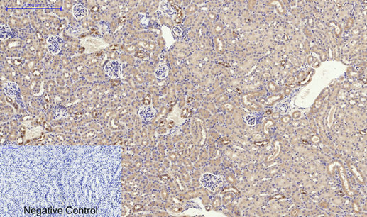
- Immunohistochemical analysis of paraffin-embedded Mouse-kidney tissue. 1,PPAR-α Polyclonal Antibody was diluted at 1:200(4°C,overnight). 2, Sodium citrate pH 6.0 was used for antibody retrieval(>98°C,20min). 3,Secondary antibody was diluted at 1:200(room tempeRature, 30min). Negative control was used by secondary antibody only.
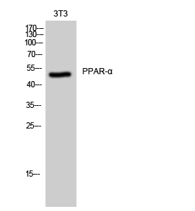
- Western Blot analysis of 3T3 cells using PPAR-α Polyclonal Antibody cells nucleus extracted by Minute TM Cytoplasmic and Nuclear Fractionation kit (SC-003,Inventbiotech,MN,USA).

- Immunofluorescence analysis of HeLa cells, using PPAR-alpha Antibody. The picture on the right is blocked with the synthesized peptide.
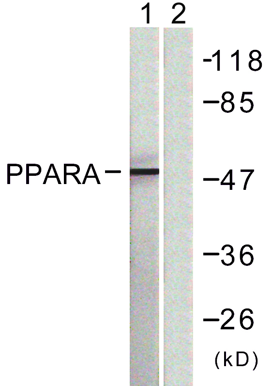
- Western blot analysis of lysates from NIH/3T3 cells, using PPAR-alpha Antibody. The lane on the right is blocked with the synthesized peptide.


