


Catalog: YP1128
Size
Price
Status
Qty.
200μL
$600.00
In stock
0
100μL
$340.00
In stock
0
50μL
$190.00
In stock
0
Add to cart


Collected
Collect
Main Information
Target
AS160
Host Species
Rabbit
Reactivity
Human, Mouse
Applications
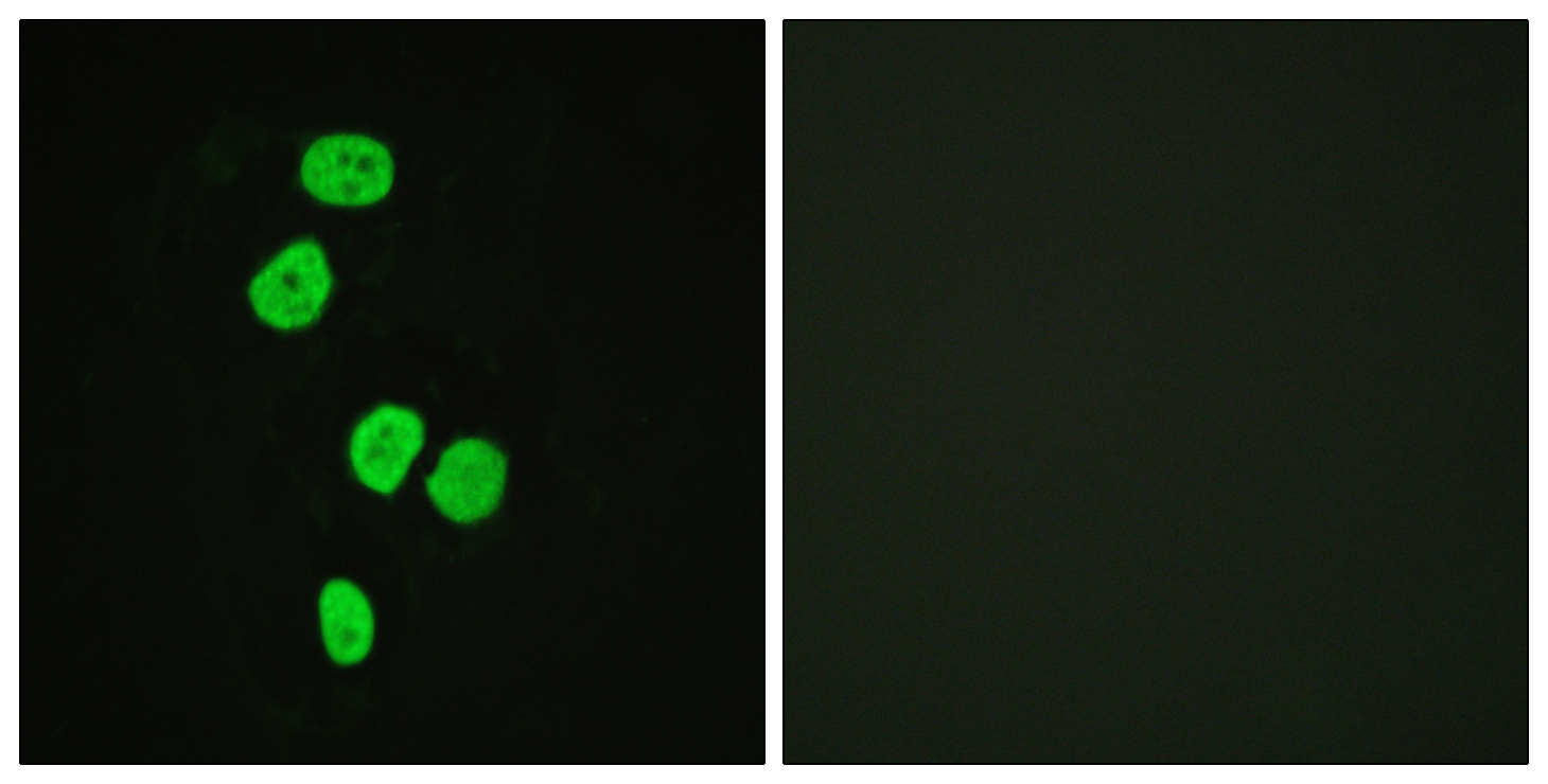
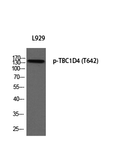
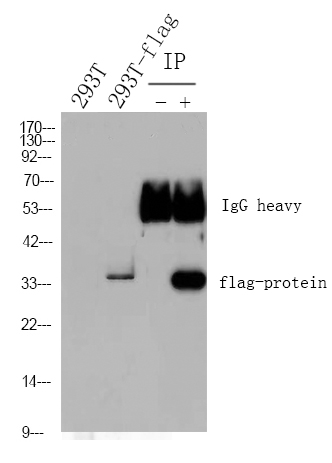
WB, IHC, IF, ELISA
MW
150kD (Observed)
Conjugate/Modification
Phospho
Detailed Information
Recommended Dilution Ratio
WB 1:500-2000; IHC 1:100-1:300; IF 1:200-1:1000; ELISA 1:5000; Not yet tested in other applications.
Formulation
Liquid in PBS containing 50% glycerol, 0.5% BSA and 0.02% sodium azide.
Specificity
Phospho-TBC1D4 (T642) Polyclonal Antibody detects endogenous levels of TBC1D4 protein only when phosphorylated at T642.The name of modified sites may be influenced by many factors, such as species (the modified site was not originally found in human samples) and the change of protein sequence (the previous protein sequence is incomplete, and the protein sequence may be prolonged with the development of protein sequencing technology). When naming, we will use the "numbers" in historical reference to keep the sites consistent with the reports. The antibody binds to the following modification sequence (lowercase letters are modification sites):AHtFS
Purification
The antibody was affinity-purified from rabbit antiserum by affinity-chromatography using epitope-specific immunogen.
Storage
-15°C to -25°C/1 year(Do not lower than -25°C)
Concentration
1 mg/ml
MW(Observed)
150kD
Modification
Phospho
Clonality
Polyclonal
Isotype
IgG
Related Products
Primary Antibodies
TBC1D4 Rabbit pAb
YT4560
More→
Primary Antibodies
TBC1D4 (Phospho Thr642) Rabbit pAb
YP1128
More→
Secondary Antibodies
Goat Anti Mouse IgG(H+L) (HRP)
RS0001
More→
Secondary Antibodies

Goat Anti Rabbit IgG(H+L) (HRP)
RS0002
More→
Primary Antibodies
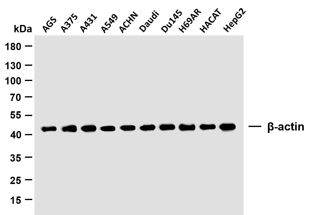
β-actin (PTR2364) Mouse mAb
YM3028
More→
Primary Antibodies
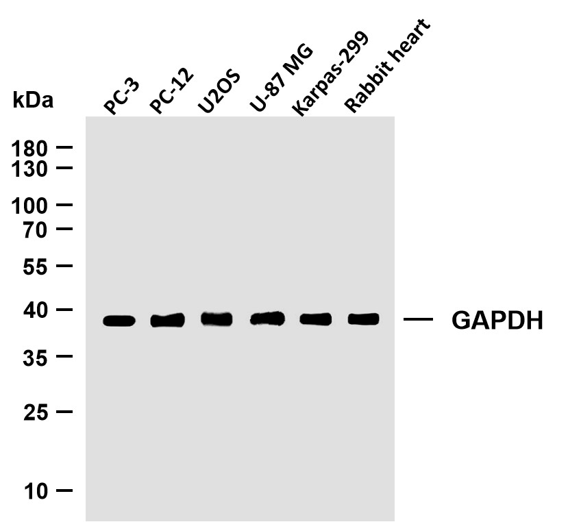
GAPDH (PTR2304) Mouse mAb
YM3029
More→
Antigen&Target Information
Immunogen:
The antiserum was produced against synthesized peptide derived from human AS160 around the phosphorylation site of Thr642. AA range:611-660
show all
Specificity:
Phospho-TBC1D4 (T642) Polyclonal Antibody detects endogenous levels of TBC1D4 protein only when phosphorylated at T642.The name of modified sites may be influenced by many factors, such as species (the modified site was not originally found in human samples) and the change of protein sequence (the previous protein sequence is incomplete, and the protein sequence may be prolonged with the development of protein sequencing technology). When naming, we will use the "numbers" in historical reference to keep the sites consistent with the reports. The antibody binds to the following modification sequence (lowercase letters are modification sites):AHtFS
show all
Gene Name:
TBC1D4 AS160 KIAA0603
show all
Protein Name:
TBC1 domain family member 4
show all
Other Name:
TBC1D4 ;
AS160 ;
KIAA0603 ;
TBC1 domain family member 4 ;
Akt substrate of 160 kDa ;
AS160
AS160 ;
KIAA0603 ;
TBC1 domain family member 4 ;
Akt substrate of 160 kDa ;
AS160
show all
Background:
This gene is a member of the Tre-2/BUB2/CDC16 domain family. The protein encoded by this gene is a Rab-GTPase-activating protein, and contains two phopshotyrosine-binding domains (PTB1 and PTB2), a calmodulin-binding domain (CBD), a Rab-GTPase domain, and multiple AKT phosphomotifs. This protein is thought to play an important role in glucose homeostasis by regulating the insulin-dependent trafficking of the glucose transporter 4 (GLUT4), important for removing glucose from the bloodstream into skeletal muscle and fat tissues. Reduced expression of this gene results in an increase in GLUT4 levels at the plasma membrane, suggesting that this protein is important in intracellular retention of GLUT4 under basal conditions. When exposed to insulin, this protein is phosphorylated, dissociates from GLUT4 vesicles, resulting in increased GLUT4 at the cell surface, and enhanced glucose transport. Ph
show all
Function:
Disease:May be involved in atopic dermatitis (AD).,Function:May act as a GTPase-activating protein for RAB2A, RAB8A, RAB10 and RAB14. Isoform 2 promotes insulin-induced glucose transporter SLC2A4/GLUT4 translocation at the plasma membrane, thus increasing glucose uptake.,PTM:Insulin-stimulated phosphorylation is required for SLC2A4/GLUT4 translocation.,PTM:Phosphorylated by AKT1; insulin-induced.,PTM:Physiological hyperinsulinemia increases phosphorylation in skeletal muscle. Insulin-stimulated phosphorylation is reduced by 39% in type 2 diabetic patients.,similarity:Contains 1 Rab-GAP TBC domain.,similarity:Contains 2 PID domains.,subcellular location:Isoform 2 shows a cytoplasmic perinuclear localization in a myoblastic cell line in resting and insulin-stimulated cells.,tissue specificity:Widely expressed, but differential expression for isoforms 1 and 2, with highest overall expression of isoform 2 in most tissues. Isoform 1 is highly expressed in skeletal muscle and heart, but was not detectable in the liver nor in adipose tissue. Isoform 2 strongly expressed in adrenal and thyroid gland, and also in lung, kidney, colon, brain and adipose tissue. Moderate isoform 2 expression in skeletal muscle. Expressed in pancreatic Langerhans islets, including beta cells (at protein level). Expression is decreased by twofold in pancreatic islets in type 2 diabetes patients compared to control subjects.,
show all
Cellular Localization:
Cytoplasm . Isoform 2 shows a cytoplasmic perinuclear localization in a myoblastic cell line in resting and insulin-stimulated cells.
show all
Tissue Expression:
Widely expressed. Isoform 2 is the highest overexpressed in most tissues. Isoform 1 is highly expressed in skeletal muscle and heart, but was not detectable in the liver nor in adipose tissue. Isoform 2 is strongly expressed in adrenal and thyroid gland, and also in lung, kidney, colon, brain and adipose tissue. Isoform 2 is moderately expressed in skeletal muscle. Expressed in pancreatic Langerhans islets, including beta cells (at protein level). Expression is decreased by twofold in pancreatic islets in type 2 diabetes patients compared to control subjects. Up-regulated in T-cells from patients with atopic dermatitis.
show all
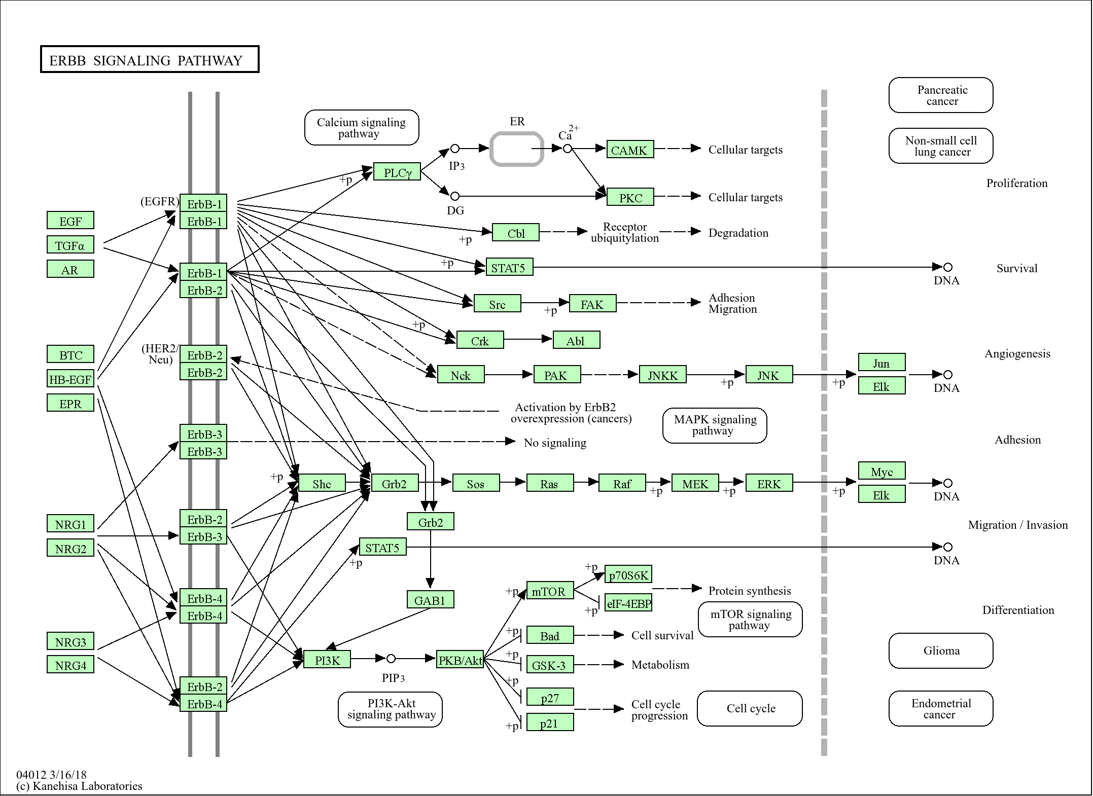
Research Areas:
>>Thyroid hormone signaling pathway ;
>>Insulin resistance ;
>>Diabetic cardiomyopathy
>>Insulin resistance ;
>>Diabetic cardiomyopathy
show all
Reference Citation({{totalcount}})
Catalog: YP1128
Size
Price
Status
Qty.
200μL
$600.00
In stock
0
100μL
$340.00
In stock
0
50μL
$190.00
In stock
0
Add to cart
Collected
Collect
Recently Viewed Products
Clear allPRODUCTS
CUSTOMIZED
ABOUT US


Toggle night Mode
{{pinfoXq.title || ''}}
Catalog: {{pinfoXq.catalog || ''}}
Filter:
All
{{item.name}}
{{pinfo.title}}
-{{pinfo.catalog}}
Main Information
Target
{{pinfo.target}}
Reactivity
{{pinfo.react}}
Applications
{{pinfo.applicat}}
Conjugate/Modification
{{pinfo.coupling}}/{{pinfo.modific}}
MW (kDa)
{{pinfo.mwcalc}}
Host Species
{{pinfo.hostspec}}
Isotype
{{pinfo.isotype}}
Product {{index}}/{{pcount}}
Prev
Next
{{pvTitle}}
Scroll wheel zooms the picture
{{pvDescr}}