


Catalog: YP0511
Size
Price
Status
Qty.
200μL
$600.00
In stock
0
100μL
$340.00
In stock
0
50μL
$190.00
In stock
0
Add to cart


Collected
Collect
Main Information
Target
Annexin II
Host Species
Rabbit
Reactivity
Human, Mouse, Rat
Applications
WB, ELISA
MW
38kD (Observed)
Conjugate/Modification
Phospho
Detailed Information
Recommended Dilution Ratio
WB 1:500-1:2000; ELISA 1:10000; Not yet tested in other applications.
Formulation
Liquid in PBS containing 50% glycerol, 0.5% BSA and 0.02% sodium azide.
Specificity
Phospho-Annexin II (S26) Polyclonal Antibody detects endogenous levels of Annexin II protein only when phosphorylated at S26.The name of modified sites may be influenced by many factors, such as species (the modified site was not originally found in human samples) and the change of protein sequence (the previous protein sequence is incomplete, and the protein sequence may be prolonged with the development of protein sequencing technology). When naming, we will use the "numbers" in historical reference to keep the sites consistent with the reports. The antibody binds to the following modification sequence (lowercase letters are modification sites):YGsVK
Purification
The antibody was affinity-purified from rabbit antiserum by affinity-chromatography using epitope-specific immunogen.
Storage
-15°C to -25°C/1 year(Do not lower than -25°C)
Concentration
1 mg/ml
MW(Observed)
38kD
Modification
Phospho
Clonality
Polyclonal
Isotype
IgG
Related Products
Secondary Antibodies
Goat Anti Mouse IgG(H+L) (HRP)
RS0001
More→
Secondary Antibodies

Goat Anti Rabbit IgG(H+L) (HRP)
RS0002
More→
Primary Antibodies
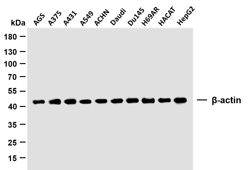
β-actin (PTR2364) Mouse mAb
YM3028
More→
Primary Antibodies
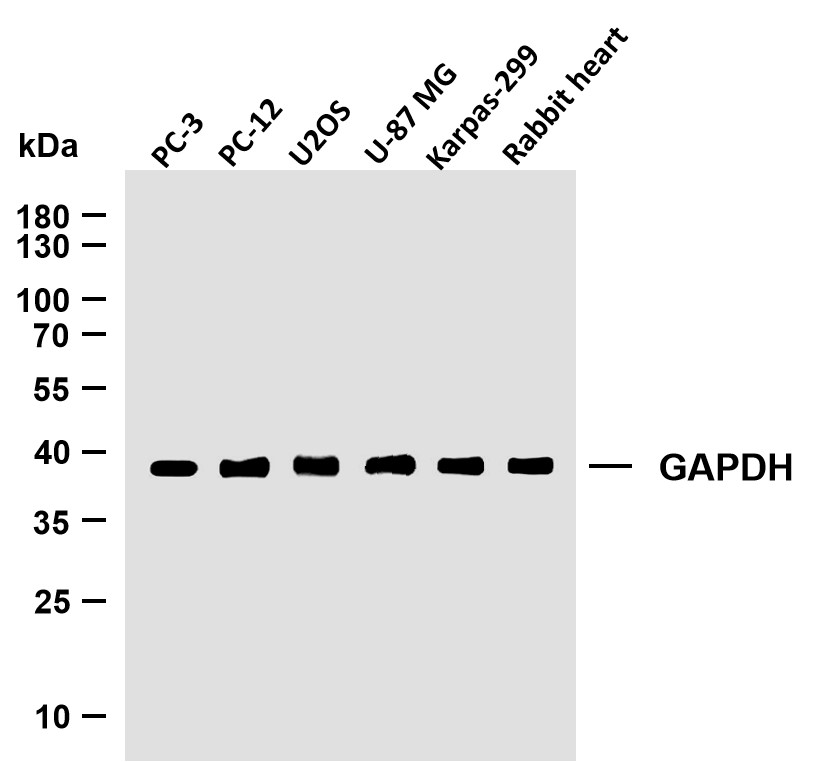
GAPDH (PTR2304) Mouse mAb
YM3029
More→
Antigen&Target Information
Immunogen:
Synthesized phospho-peptide around the phosphorylation site of human Annexin II (phospho Ser26)
show all
Specificity:
Phospho-Annexin II (S26) Polyclonal Antibody detects endogenous levels of Annexin II protein only when phosphorylated at S26.The name of modified sites may be influenced by many factors, such as species (the modified site was not originally found in human samples) and the change of protein sequence (the previous protein sequence is incomplete, and the protein sequence may be prolonged with the development of protein sequencing technology). When naming, we will use the "numbers" in historical reference to keep the sites consistent with the reports. The antibody binds to the following modification sequence (lowercase letters are modification sites):YGsVK
show all
Gene Name:
ANXA2
show all
Protein Name:
Annexin A2
show all
Other Name:
ANXA2 ;
ANX2 ;
ANX2L4 ;
CAL1H ;
LPC2D ;
Annexin A2 ;
Annexin II ;
Annexin-2 ;
Calpactin I heavy chain ;
Calpactin-1 heavy chain ;
Chromobindin-8 ;
Lipocortin II ;
Placental anticoagulant protein IV ;
PAP-IV ;
Protein I ;
p36
ANX2 ;
ANX2L4 ;
CAL1H ;
LPC2D ;
Annexin A2 ;
Annexin II ;
Annexin-2 ;
Calpactin I heavy chain ;
Calpactin-1 heavy chain ;
Chromobindin-8 ;
Lipocortin II ;
Placental anticoagulant protein IV ;
PAP-IV ;
Protein I ;
p36
show all
Database Link:
Background:
This gene encodes a member of the annexin family. Members of this calcium-dependent phospholipid-binding protein family play a role in the regulation of cellular growth and in signal transduction pathways. This protein functions as an autocrine factor which heightens osteoclast formation and bone resorption. This gene has three pseudogenes located on chromosomes 4, 9 and 10, respectively. Multiple alternatively spliced transcript variants encoding different isoforms have been found for this gene. [provided by RefSeq, Jul 2008],
show all
Function:
Domain:A pair of annexin repeats may form one binding site for calcium and phospholipid.,Function:Calcium-regulated membrane-binding protein whose affinity for calcium is greatly enhanced by anionic phospholipids. It binds two calcium ions with high affinity. May be involved in heat-stress response.,miscellaneous:It may cross-link plasma membrane phospholipids with actin and the cytoskeleton and be involved with exocytosis.,online information:Red velvet - Issue 86 of September 2007,PTM:Phosphorylation of Tyr-24 enhances heat stress-induced translocation to the cell surface.,similarity:Belongs to the annexin family.,similarity:Contains 4 annexin repeats.,subcellular location:In the lamina beneath the plasma membrane. Identified by mass spectrometry in melanosome fractions from stage I to stage IV. Translocated from the cytoplasm to the cell surface through a Golgi-independent mechanism.,subunit:Heterotetramer containing 2 light chains of S100A10/p11 and 2 heavy chains of ANXA2/p36. Interacts with ATP1B1 and DYSF.,
show all
Cellular Localization:
Secreted, extracellular space, extracellular matrix, basement membrane . Melanosome . In the lamina beneath the plasma membrane. Identified by mass spectrometry in melanosome fractions from stage I to stage IV. Translocated from the cytoplasm to the cell surface through a Golgi-independent mechanism.
show all
Tissue Expression:
Brain,Colon,Colon adenocarcinoma,Epithelium,Osteosarcoma,Pancreas,Placenta,Prostate,Skin,Te
show all
Research Areas:
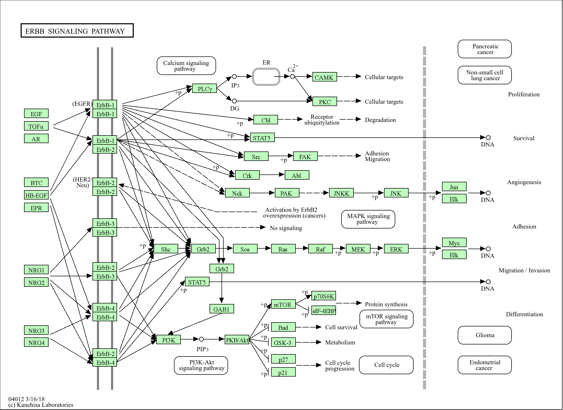
>>Salmonella infection
show all
Reference Citation({{totalcount}})
Catalog: YP0511
Size
Price
Status
Qty.
200μL
$600.00
In stock
0
100μL
$340.00
In stock
0
50μL
$190.00
In stock
0
Add to cart
Collected
Collect
Recently Viewed Products
Clear allPRODUCTS
CUSTOMIZED
ABOUT US


Toggle night Mode
{{pinfoXq.title || ''}}
Catalog: {{pinfoXq.catalog || ''}}
Filter:
All
{{item.name}}
{{pinfo.title}}
-{{pinfo.catalog}}
Main Information
Target
{{pinfo.target}}
Reactivity
{{pinfo.react}}
Applications
{{pinfo.applicat}}
Conjugate/Modification
{{pinfo.coupling}}/{{pinfo.modific}}
MW (kDa)
{{pinfo.mwcalc}}
Host Species
{{pinfo.hostspec}}
Isotype
{{pinfo.isotype}}
Product {{index}}/{{pcount}}
Prev
Next
{{pvTitle}}
Scroll wheel zooms the picture
{{pvDescr}}