


Catalog: YP0134
Size
Price
Status
Qty.
200μL
$600.00
In stock
0
100μL
$340.00
In stock
0
50μL
$190.00
In stock
0
Add to cart


Collected
Collect
Main Information
Target
HSP27
Host Species
Rabbit
Reactivity
Human, Monkey
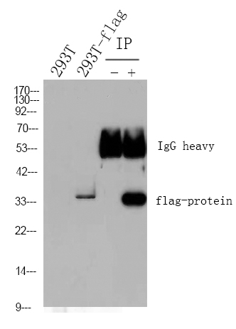
Applications
WB, IHC, IF, ELISA
MW
26kD (Observed)
Conjugate/Modification
Phospho
Detailed Information
Recommended Dilution Ratio
WB 1:500-1:2000; IHC 1:100-1:300; ELISA 1:20000; IF 1:50-200
Formulation
Liquid in PBS containing 50% glycerol, 0.5% BSA and 0.02% sodium azide.
Specificity
Phospho-HSP27 (S15) Polyclonal Antibody detects endogenous levels of HSP27 protein only when phosphorylated at S15.The name of modified sites may be influenced by many factors, such as species (the modified site was not originally found in human samples) and the change of protein sequence (the previous protein sequence is incomplete, and the protein sequence may be prolonged with the development of protein sequencing technology). When naming, we will use the "numbers" in historical reference to keep the sites consistent with the reports. The antibody binds to the following modification sequence (lowercase letters are modification sites):GPsWD
Purification
The antibody was affinity-purified from rabbit antiserum by affinity-chromatography using epitope-specific immunogen.
Storage
-15°C to -25°C/1 year(Do not lower than -25°C)
Concentration
1 mg/ml
MW(Observed)
26kD
Modification
Phospho
Clonality
Polyclonal
Isotype
IgG
Related Products
Secondary Antibodies
Goat Anti Mouse IgG(H+L) (HRP)
RS0001
More→
Secondary Antibodies

Goat Anti Rabbit IgG(H+L) (HRP)
RS0002
More→
Primary Antibodies
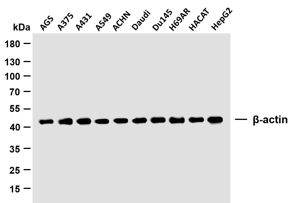
β-actin (PTR2364) Mouse mAb
YM3028
More→
Primary Antibodies
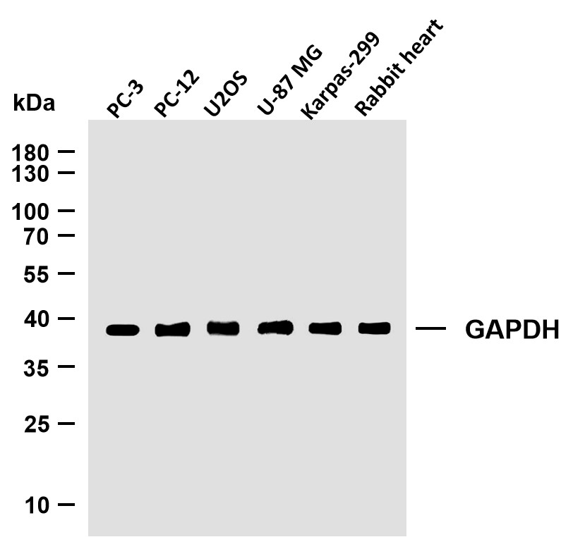
GAPDH (PTR2304) Mouse mAb
YM3029
More→
Antigen&Target Information
Immunogen:
The antiserum was produced against synthesized peptide derived from human HSP27 around the phosphorylation site of Ser15. AA range:5-54
show all
Specificity:
Phospho-HSP27 (S15) Polyclonal Antibody detects endogenous levels of HSP27 protein only when phosphorylated at S15.The name of modified sites may be influenced by many factors, such as species (the modified site was not originally found in human samples) and the change of protein sequence (the previous protein sequence is incomplete, and the protein sequence may be prolonged with the development of protein sequencing technology). When naming, we will use the "numbers" in historical reference to keep the sites consistent with the reports. The antibody binds to the following modification sequence (lowercase letters are modification sites):GPsWD
show all
Gene Name:
HSPB1 HSP27 HSP28
show all
Protein Name:
Heat shock protein beta-1
show all
Other Name:
HSPB1 ;
HSP27 ;
HSP28 ;
Heat shock protein beta-1 ;
HspB1 ;
28 kDa heat shock protein ;
Estrogen-regulated 24 kDa protein ;
Heat shock 27 kDa protein ;
HSP 27 ;
Stress-responsive protein 27 ;
SRP27
HSP27 ;
HSP28 ;
Heat shock protein beta-1 ;
HspB1 ;
28 kDa heat shock protein ;
Estrogen-regulated 24 kDa protein ;
Heat shock 27 kDa protein ;
HSP 27 ;
Stress-responsive protein 27 ;
SRP27
show all
Background:
The protein encoded by this gene is induced by environmental stress and developmental changes. The encoded protein is involved in stress resistance and actin organization and translocates from the cytoplasm to the nucleus upon stress induction. Defects in this gene are a cause of Charcot-Marie-Tooth disease type 2F (CMT2F) and distal hereditary motor neuropathy (dHMN). [provided by RefSeq, Oct 2008],
show all
Function:
Disease:Defects in HSPB1 are a cause of distal hereditary motor neuronopathy type 2B (HMN2B) [MIM:608634]. Distal hereditary motor neuronopathies constitute a heterogeneous group of neuromuscular disorders caused by selective impairment of motor neurons in the anterior horn of the spinal cord, without sensory deficit in the posterior horn. The overall clinical picture consists of a classical distal muscular atrophy syndrome in the legs without clinical sensory loss. The disease starts with weakness and wasting of distal muscles of the anterior tibial and peroneal compartments of the legs. Later on, weakness and atrophy may expand to the proximal muscles of the lower limbs and/or to the distal upper limbs.,Disease:Defects in HSPB1 are the cause of Charcot-Marie-Tooth disease type 2F (CMT2F) [MIM:606595]. CMT2F is a form of Charcot-Marie-Tooth disease, the most common inherited disorder of the peripheral nervous system. Charcot-Marie-Tooth disease is classified in two main groups on the basis of electrophysiologic properties and histopathology: primary peripheral demyelinating neuropathy or CMT1, and primary peripheral axonal neuropathy or CMT2. Neuropathies of the CMT2 group are characterized by signs of axonal regeneration in the absence of obvious myelin alterations, normal or slightly reduced nerve conduction velocities, and progressive distal muscle weakness and atrophy. Nerve conduction velocities are normal or slightly reduced. CMT2F onset is between 15 and 25 years with muscle weakness and atrophy usually beginning in feet and legs (peroneal distribution). Upper limb involvement occurs later. CMT2F inheritance is autosomal dominant.,Function:Involved in stress resistance and actin organization.,induction:Expressed in response to environmental stresses such as heat shock, or estrogen stimulation in MCF-7 cells.,PTM:Phosphorylated in MCF-7 cells on exposure to protein kinase C activators and heat shock.,similarity:Belongs to the small heat shock protein (HSP20) family.,subcellular location:Cytoplasmic in interphase cells. Colocalizes with mitotic spindles in mitotic cells. Translocates to the nucleus during heat shock.,subunit:Interacts with TGFB1I1 (By similarity). Associates with alpha- and beta-tubulin, microtubules and CRYAB. Interacts with HSPB8 and HSPBAP1.,tissue specificity:Detected in all tissues tested: skeletal muscle, heart, aorta, large intestine, small intestine, stomach, esophagus, bladder, adrenal gland, thyroid, pancreas, testis, adipose tissue, kidney, liver, spleen, cerebral cortex, blood serum and cerebrospinal fluid. Highest levels are found in the heart and in tissues composed of striated and smooth muscle.,
show all
Cellular Localization:
Cytoplasm . Nucleus . Cytoplasm, cytoskeleton, spindle . Cytoplasmic in interphase cells. Colocalizes with mitotic spindles in mitotic cells. Translocates to the nucleus during heat shock and resides in sub-nuclear structures known as SC35 speckles or nuclear splicing speckles. .
show all
Tissue Expression:
Detected in all tissues tested: skeletal muscle, heart, aorta, large intestine, small intestine, stomach, esophagus, bladder, adrenal gland, thyroid, pancreas, testis, adipose tissue, kidney, liver, spleen, cerebral cortex, blood serum and cerebrospinal fluid. Highest levels are found in the heart and in tissues composed of striated and smooth muscle.
show all
Research Areas:
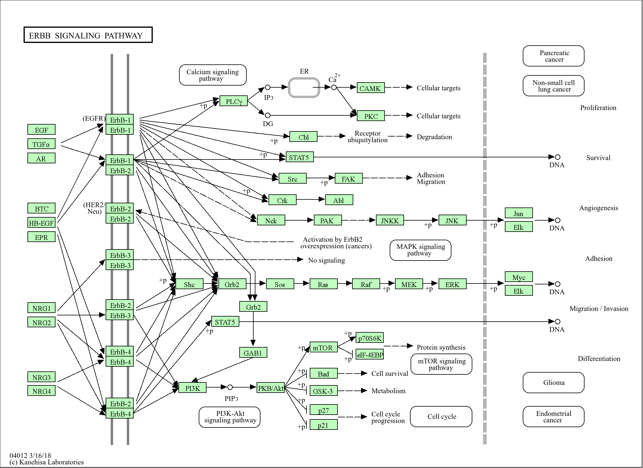
>>MAPK signaling pathway ;
>>VEGF signaling pathway ;
>>Amoebiasis
>>VEGF signaling pathway ;
>>Amoebiasis
show all
Signaling Pathway
Reference Citation({{totalcount}})
Catalog: YP0134
Size
Price
Status
Qty.
200μL
$600.00
In stock
0
100μL
$340.00
In stock
0
50μL
$190.00
In stock
0
Add to cart
Collected
Collect
Recently Viewed Products
Clear allPRODUCTS
CUSTOMIZED
ABOUT US


Toggle night Mode
{{pinfoXq.title || ''}}
Catalog: {{pinfoXq.catalog || ''}}
Filter:
All
{{item.name}}
{{pinfo.title}}
-{{pinfo.catalog}}
Main Information
Target
{{pinfo.target}}
Reactivity
{{pinfo.react}}
Applications
{{pinfo.applicat}}
Conjugate/Modification
{{pinfo.coupling}}/{{pinfo.modific}}
MW (kDa)
{{pinfo.mwcalc}}
Host Species
{{pinfo.hostspec}}
Isotype
{{pinfo.isotype}}
Product {{index}}/{{pcount}}
Prev
Next
{{pvTitle}}
Scroll wheel zooms the picture
{{pvDescr}}