


Catalog: YN6145
Size
Price
Status
Qty.
200μL
$450.00
In stock
0
100μL
$280.00
In stock
0
40μL
$160.00
In stock
0
Add to cart


Collected
Collect
Main Information
Target
MAP1S
Host Species
Rabbit
Reactivity
Human, Mouse, Rat
Applications
WB
MW
116kD (Calculated)
Conjugate/Modification
Unmodified
Detailed Information
Recommended Dilution Ratio
WB 1:500-2000
Formulation
Liquid in PBS containing 50% glycerol, 0.5% BSA and 0.02% sodium azide.
Specificity
This antibody detects endogenous levels of MAP1S at Human, Mouse,Rat
Purification
The antibody was affinity-purified from rabbit antiserum by affinity-chromatography using epitope-specific immunogen.
Storage
-15°C to -25°C/1 year(Do not lower than -25°C)
Concentration
1 mg/ml
MW(Calculated)
116kD
Modification
Unmodified
Clonality
Polyclonal
Isotype
IgG
Related Products
Secondary Antibodies
Goat Anti Mouse IgG(H+L) (HRP)
RS0001
More→
Secondary Antibodies

Goat Anti Rabbit IgG(H+L) (HRP)
RS0002
More→
Primary Antibodies
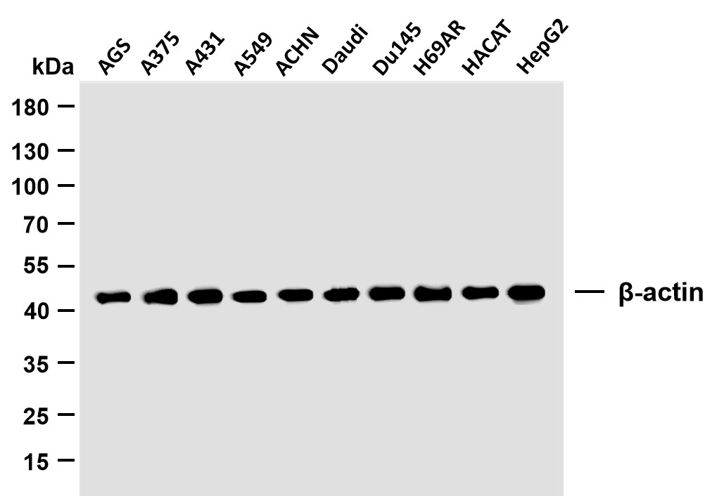
β-actin (PTR2364) Mouse mAb
YM3028
More→
Primary Antibodies
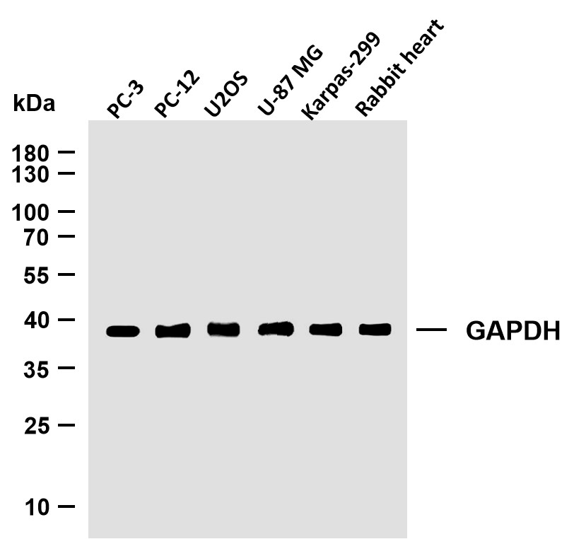
GAPDH (PTR2304) Mouse mAb
YM3029
More→
Antigen&Target Information
Immunogen:
Synthesized peptide derived from human MAP1S
show all
Specificity:
This antibody detects endogenous levels of MAP1S at Human, Mouse,Rat
show all
Gene Name:
MAP1S BPY2IP1 C19orf5 MAP8 VCY2IP1
show all
Protein Name:
Microtubule-associated protein 1S (MAP-1S) (BPY2-interacting protein 1) (Microtubule-associated protein 8) (Variable charge Y chromosome 2-interacting protein 1) (VCY2-interacting protein 1) (VCY2IP-1) [Cleaved into: MAP1S heavy chain; MAP1S light chain]
show all
Database Link:
Function:
Microtubule-associated protein that mediates aggregation of mitochondria resulting in cell death and genomic destruction (MAGD). Plays a role in anchoring the microtubule organizing center to the centrosomes. Binds to DNA. Plays a role in apoptosis. Involved in the formation of microtubule bundles (By similarity).
show all
Cellular Localization:
Nucleus. Cytoplasm, cytosol. Cytoplasm, cytoskeleton . Cytoplasm, cytoskeleton, spindle . Detected in filopodia-like protrusions and synapses (By similarity). Detected in perinuclear punctate network corresponding to mitochondrial aggregates and in the nucleus in cells exhibiting apoptosis. Associated specifically with microtubules stabilized by paclitaxel and colocalizes with RASSF1 isoform A. In interphase cells, shows a diffuse cytoplasmic staining with partial localization to the microtubules. During the different stages of mitosis detected at the spindle microtubules. .
show all
Reference Citation({{totalcount}})
Catalog: YN6145
Size
Price
Status
Qty.
200μL
$450.00
In stock
0
100μL
$280.00
In stock
0
40μL
$160.00
In stock
0
Add to cart
Collected
Collect
Recently Viewed Products
Clear allPRODUCTS
CUSTOMIZED
ABOUT US


Toggle night Mode
{{pinfoXq.title || ''}}
Catalog: {{pinfoXq.catalog || ''}}
Filter:
All
{{item.name}}
{{pinfo.title}}
-{{pinfo.catalog}}
Main Information
Target
{{pinfo.target}}
Reactivity
{{pinfo.react}}
Applications
{{pinfo.applicat}}
Conjugate/Modification
{{pinfo.coupling}}/{{pinfo.modific}}
MW (kDa)
{{pinfo.mwcalc}}
Host Species
{{pinfo.hostspec}}
Isotype
{{pinfo.isotype}}
Product {{index}}/{{pcount}}
Prev
Next
{{pvTitle}}
Scroll wheel zooms the picture
{{pvDescr}}