


Catalog: YN2221
Size
Price
Status
Qty.
200μL
$450.00
In stock
0
100μL
$280.00
In stock
0
40μL
$150.00
In stock
0
Add to cart


Collected
Collect
Main Information
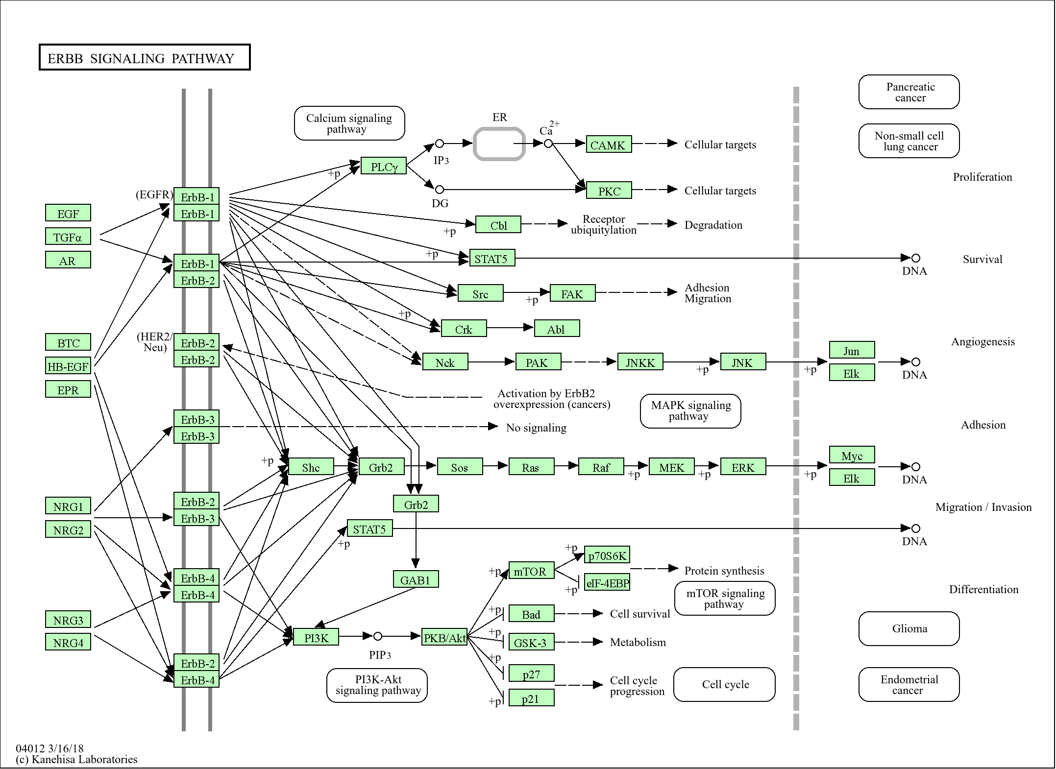
Target
BGH3
Host Species
Rabbit
Reactivity
Human, Mouse
Applications
WB, ELISA
MW
75kD (Observed)
Conjugate/Modification
Unmodified
Detailed Information
Recommended Dilution Ratio
WB 1:500-2000; ELISA 1:5000-20000
Formulation
Liquid in PBS containing 50% glycerol,0.5% BSA and 0.02% sodium azide.
Specificity
BGH3 Polyclonal Antibody detects endogenous levels of protein.
Purification
The antibody was affinity-purified from rabbit antiserum by affinity-chromatography using epitope-specific immunogen.
Storage
-15°C to -25°C/1 year(Do not lower than -25°C)
Concentration
1 mg/ml
MW(Observed)
75kD
Modification
Unmodified
Clonality
Polyclonal
Isotype
IgG
Related Products
Secondary Antibodies
Goat Anti Mouse IgG(H+L) (HRP)
RS0001
More→
Secondary Antibodies

Goat Anti Rabbit IgG(H+L) (HRP)
RS0002
More→
Primary Antibodies
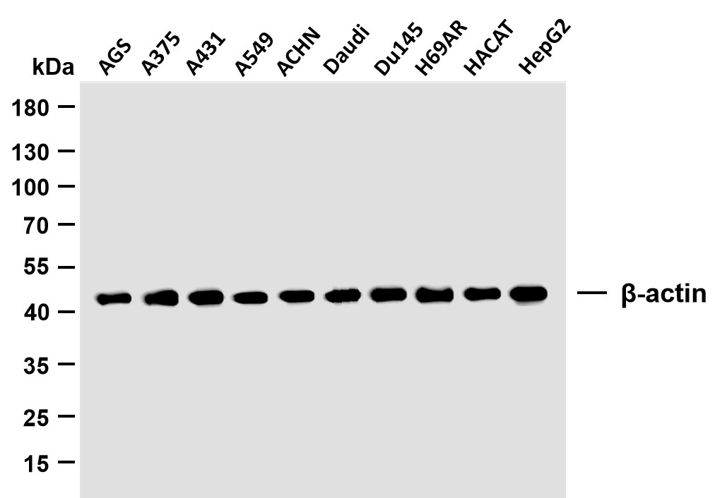
β-actin (PTR2364) Mouse mAb
YM3028
More→
Primary Antibodies
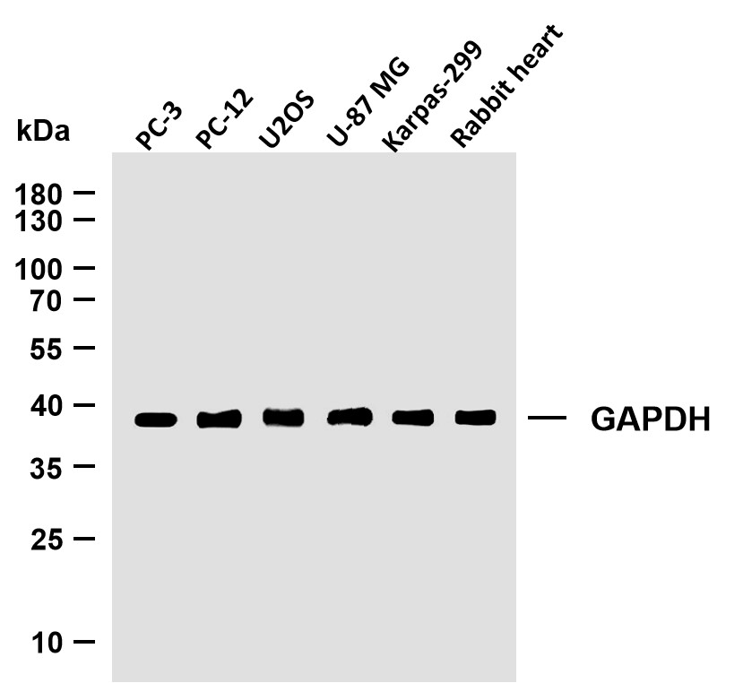
GAPDH (PTR2304) Mouse mAb
YM3029
More→
Antigen&Target Information
Immunogen:
Synthesized peptide derived from human protein . at AA range: 230-310
show all
Specificity:
BGH3 Polyclonal Antibody detects endogenous levels of protein.
show all
Gene Name:
TGFBI BIGH3
show all
Protein Name:
Transforming growth factor-beta-induced protein ig-h3 (Beta ig-h3) (Kerato-epithelin) (RGD-containing collagen-associated protein) (RGD-CAP)
show all
Background:
This gene encodes an RGD-containing protein that binds to type I, II and IV collagens. The RGD motif is found in many extracellular matrix proteins modulating cell adhesion and serves as a ligand recognition sequence for several integrins. This protein plays a role in cell-collagen interactions and may be involved in endochondrial bone formation in cartilage. The protein is induced by transforming growth factor-beta and acts to inhibit cell adhesion. Mutations in this gene are associated with multiple types of corneal dystrophy. [provided by RefSeq, Jul 2008],
show all
Function:
Disease:Defects in TGFBI are a cause of corneal dystrophy Thiel-Behnke type (CDTB) [MIM:602082]; also known as corneal dystrophy of Bowman layer type 2 (CDB2).,Disease:Defects in TGFBI are the cause of Avellino corneal dystrophy (ACD) [MIM:607541]. ACD could be considered a variant of granular dystrophy with a significant amyloidogenic tendency. Inheritance is autosomal dominant.,Disease:Defects in TGFBI are the cause of corneal dystrophy Groenouw type 1 (CDGG1) [MIM:121900]; also known as corneal dystrophy granular type. Inheritance is autosomal dominant. Corneal dystrophies show progressive opacification of the cornea leading to severe visual handicap.,Disease:Defects in TGFBI are the cause of corneal dystrophy lattice type 1 (CDL1) [MIM:122200]. Inheritance is autosomal dominant.,Disease:Defects in TGFBI are the cause of epithelial basement membrane corneal dystrophy (EBMD) [MIM:121820]; also known as Cogan corneal dystrophy or map-dot-fingerprint type corneal dystrophy. EBMD is a bilateral anterior corneal dystrophy characterized by grayish epithelial fingerprint lines, geographic map-like lines, and dots (or microcysts) on slit-lamp examination. Pathologic studies show abnormal, redundant basement membrane and intraepithelial lacunae filled with cellular debris. Although this disorder usually is not considered to be inherited, families with autosomal dominant inheritance have been identified.,Disease:Defects in TGFBI are the cause of lattice corneal dystrophy type 3A (CDL3A) [MIM:608471]. CDL3A clinically resembles to lattice corneal dystrophy type 3, but differs in that its age of onset is 70 to 90 years. It has an autosomal dominant inheritance pattern.,Disease:Defects in TGFBI are the cause of Reis-Buecklers corneal dystrophy (CDRB) [MIM:608470]; also known as corneal dystrophy of Bowman layer type 1 (CDB1).,Function:Binds to type I, II, and IV collagens. This adhesion protein may play an important role in cell-collagen interactions. In cartilage, may be involved in endochondral bone formation.,induction:By TGF-beta.,PTM:Gamma-carboxyglutamate residues are formed by vitamin K dependent carboxylation. These residues are essential for the binding of calcium.,similarity:Contains 1 EMI domain.,similarity:Contains 4 FAS1 domains.,subcellular location:May be associated both with microfibrils and with the cell surface.,tissue specificity:Highly expressed in the corneal epithelium.,
show all
Cellular Localization:
Secreted . Secreted, extracellular space, extracellular matrix . May be associated both with microfibrils and with the cell surface (PubMed:8077289). .
show all
Reference Citation({{totalcount}})
Catalog: YN2221
Size
Price
Status
Qty.
200μL
$450.00
In stock
0
100μL
$280.00
In stock
0
40μL
$150.00
In stock
0
Add to cart
Collected
Collect
Recently Viewed Products
Clear allPRODUCTS
CUSTOMIZED
ABOUT US


Toggle night Mode
{{pinfoXq.title || ''}}
Catalog: {{pinfoXq.catalog || ''}}
Filter:
All
{{item.name}}
{{pinfo.title}}
-{{pinfo.catalog}}
Main Information
Target
{{pinfo.target}}
Reactivity
{{pinfo.react}}
Applications
{{pinfo.applicat}}
Conjugate/Modification
{{pinfo.coupling}}/{{pinfo.modific}}
MW (kDa)
{{pinfo.mwcalc}}
Host Species
{{pinfo.hostspec}}
Isotype
{{pinfo.isotype}}
Product {{index}}/{{pcount}}
Prev
Next
{{pvTitle}}
Scroll wheel zooms the picture
{{pvDescr}}