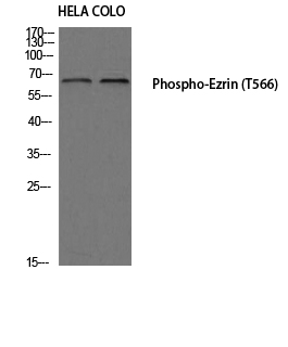
Catalog: KA1080C
Size
Price
Status
Qty.
96well
$470.00
In stock
0
Add to cart


Collected


Collect
Main Information
Reactivity
Human, Mouse, Rat
Applications
ELISA
Conjugate/Modification
Phospho
Detailed Information
Storage
2-8°C/6 months,Ship by ice bag
Modification
Phospho
Detection Method
Colorimetric
Related Products
Antigen&Target Information
Gene Name:
EZR
show all
Protein Name:
Ezrin
show all
Other Name:
EZR ;
VIL2 ;
Ezrin ;
Cytovillin ;
Villin-2 ;
p81
VIL2 ;
Ezrin ;
Cytovillin ;
Villin-2 ;
p81
show all
Database Link:
Background:
developmental stage:Very strong staining is detected in the Purkinje cell layer and in part of the molecular layer of the infant brain compared to adult brain.,function:Probably involved in connections of major cytoskeletal structures to the plasma membrane. In epithelial cells, required for the formation of microvilli and membrane ruffles on the apical pole. Along with PLEKHG6, required for normal macropinocytosis.,PTM:Phosphorylated by tyrosine-protein kinases.,similarity:Contains 1 FERM domain.,subcellular location:Localization to the apical membrane of parietal cells depends on the interaction with MPP5. Localizes to cell extensions and peripheral processes of astrocytes (By similarity). Microvillar peripheral membrane protein (cytoplasmic side).,subunit:Interacts with MPP5 (By similarity). Interacts with SLC9A3R1 and SCYL3/PACE1. Interacts with PLEKHG6. Interacts with NGX6.,tissue specificity:Expressed in cerebral cortex, basal ganglia, hippocampus, hypophysis, and optic nerve. Weakly expressed in brain stem and diencephalon. Stronger expression was detected in gray matter of frontal lobe compared to white matter (at protein level). Component of the microvilli of intestinal epithelial cells. Preferentially expressed in astrocytes of hippocampus, frontal cortex, thalamus, parahippocampal cortex, amygdala, insula, and corpus callosum. Not detected in neurons in most tissues studied.,
show all
Function:
cell morphogenesis, cytoskeleton organization, actin filament organization, cytoskeletal anchoring at plasma membrane, cell adhesion, leukocyte adhesion, establishment or maintenance of cell polarity, protein localization,regulation of cell shape, cell-cell adhesion, membrane docking, regulation of cell morphogenesis, biological adhesion,membrane to membrane docking, actin filament-based process, actin cytoskeleton organization, epithelial cell differentiation, maintenance of protein location in cell, cellular component morphogenesis, establishment or maintenance of apical/basal cell polarity, maintenance of protein location, actin filament bundle formation,maintenance of location, maintenance of location in cell, epithelium development,
show all
Cellular Localization:
Apical cell membrane ; Peripheral membrane protein ; Cytoplasmic side . Cell projection . Cell projection, microvillus membrane ; Peripheral membrane protein ; Cytoplasmic side . Cell projection, ruffle membrane ; Peripheral membrane protein ; Cytoplasmic side . Cytoplasm, cell cortex . Cytoplasm, cytoskeleton . Cell projection, microvillus . Localization to the apical membrane of parietal cells depends on the interaction with PALS1. Localizes to cell extensions and peripheral processes of astrocytes (By similarity). Microvillar peripheral membrane protein (cytoplasmic side). .
show all
Signaling Pathway
Reference Citation({{totalcount}})
Catalog: KA1080C
Size
Price
Status
Qty.
96well
$470.00
In stock
0
Add to cart


Collected


Collect
Recently Viewed Products
Clear allPRODUCTS
CUSTOMIZED
ABOUT US
Toggle night Mode
{{pinfoXq.title || ''}}
Catalog: {{pinfoXq.catalog || ''}}
Filter:
All
{{item.name}}
{{pinfo.title}}
-{{pinfo.catalog}}
Main Information
Target
{{pinfo.target}}
Reactivity
{{pinfo.react}}
Applications
{{pinfo.applicat}}
Conjugate/Modification
{{pinfo.coupling}}/{{pinfo.modific}}
MW (kDa)
{{pinfo.mwcalc}}
Host Species
{{pinfo.hostspec}}
Isotype
{{pinfo.isotype}}
Product {{index}}/{{pcount}}
Prev
Next
{{pvTitle}}
Scroll wheel zooms the picture
{{pvDescr}}

