


Catalog: YP1253
Size
Price
Status
Qty.
200μL
$600.00
In stock
0
100μL
$340.00
In stock
0
50μL
$190.00
In stock
0
Add to cart


Collected
Collect
Main Information
Target
53BP1 Phospho Ser1618
Host Species
Rabbit
Reactivity
Human, Rat
Applications
WB
MW
213kD (Observed)
Conjugate/Modification
Phospho
Detailed Information
Recommended Dilution Ratio
WB 1:1000-2000
Formulation
Liquid in PBS containing 50% glycerol, 0.5% BSA and 0.02% sodium azide.
Specificity
This antibody detects endogenous levels of Human Rat 53BP1 (phospho-Ser1618)
Purification
The antibody was affinity-purified from rabbit serum by affinity-chromatography using specific immunogen.
Storage
-15°C to -25°C/1 year(Do not lower than -25°C)
Concentration
1 mg/ml
MW(Observed)
213kD
Modification
Phospho
Clonality
Polyclonal
Isotype
IgG
Related Products
Secondary Antibodies
Goat Anti Mouse IgG(H+L) (HRP)
RS0001
More→
Secondary Antibodies

Goat Anti Rabbit IgG(H+L) (HRP)
RS0002
More→
Primary Antibodies
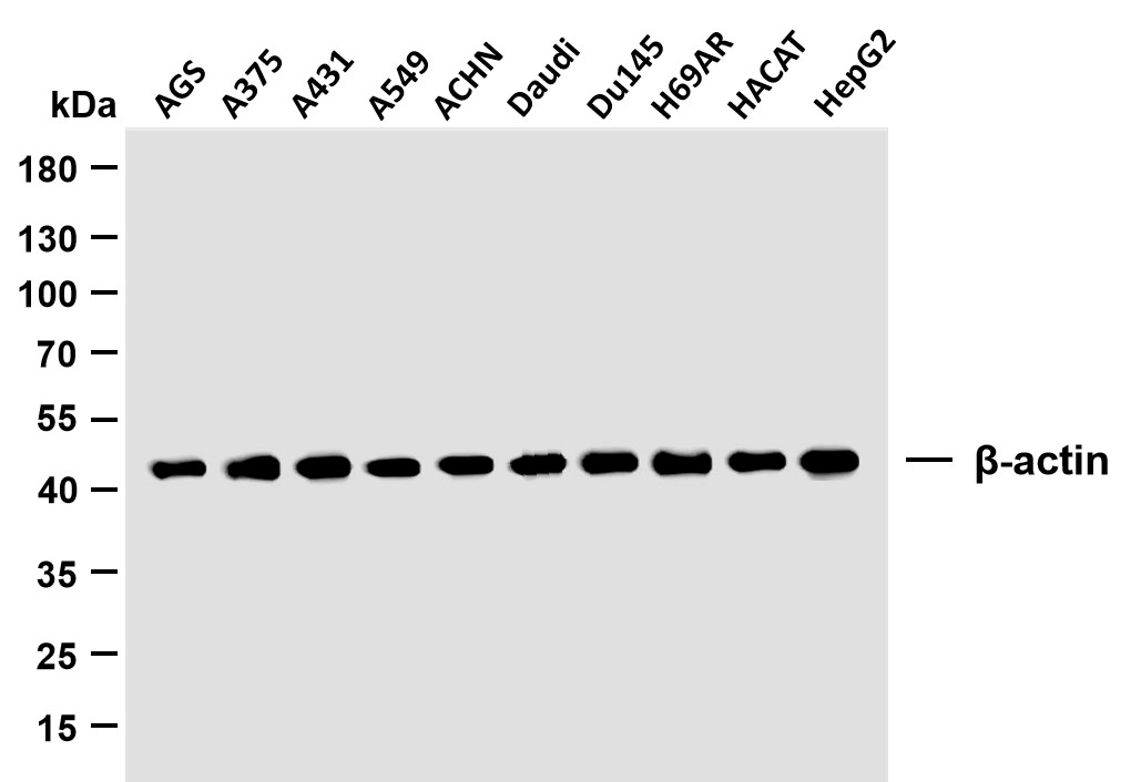
β-actin (PTR2364) Mouse mAb
YM3028
More→
Primary Antibodies
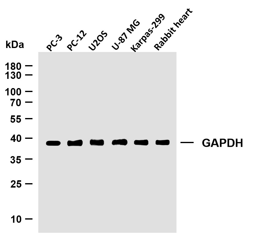
GAPDH (PTR2304) Mouse mAb
YM3029
More→
Antigen&Target Information
Immunogen:
Synthesized phosho peptide around human 53BP1 (Ser1618)
show all
Specificity:
This antibody detects endogenous levels of Human Rat 53BP1 (phospho-Ser1618)
show all
Gene Name:
TP53BP1
show all
Protein Name:
53BP1 (Ser1618)
show all
Other Name:
TP53BP1 ;
Tumor suppressor p53-binding protein 1 ;
53BP1 ;
p53-binding protein 1 ;
p53BP1
Tumor suppressor p53-binding protein 1 ;
53BP1 ;
p53-binding protein 1 ;
p53BP1
show all
Background:
This gene encodes a protein that functions in the DNA double-strand break repair pathway choice, promoting non-homologous end joining (NHEJ) pathways, and limiting homologous recombination. This protein plays multiple roles in the DNA damage response, including promoting checkpoint signaling following DNA damage, acting as a scaffold for recruitment of DNA damage response proteins to damaged chromatin, and promoting NHEJ pathways by limiting end resection following a double-strand break. These roles are also important during V(D)J recombination, class switch recombination and at unprotected telomeres. Alternative splicing results in multiple transcript variants encoding different isoforms. [provided by RefSeq, Aug 2017]
show all
Function:
Function:May have a role in checkpoint signaling during mitosis (By similarity). Enhances TP53-mediated transcriptional activation. Plays a role in the response to DNA damage.,PTM:Asymmetrically dimethylated on Arg residues by PRMT1. Methylation is required for DNA binding.,PTM:Phosphorylated at basal level in the absence of DNA damage. Hyper-phosphorylated in an ATM-dependent manner in response to DNA damage induced by ionizing radiation. Hyper-phosphorylated in an ATR-dependent manner in response to DNA damage induced by UV irradiation.,similarity:Contains 2 BRCT domains.,subcellular location:Associated with kinetochores. Both nuclear and cytoplasmic in some cells. Recruited to sites of DNA damage, such as double stand breaks. Methylation of histone H4 at 'Lys-20' is required for efficient localization to double strand breaks.,subunit:Interacts with IFI202A (By similarity). Binds to the central domain of TP53/p53. May form homo-oligomers. Interacts with DCLRE1C. Interacts with histone H2AFX and this requires phosphorylation of H2AFX on 'Ser-139'. Interacts with histone H4 that has been dimethylated at 'Lys-20'. Has low affinity for histone H4 containing monomethylated 'Lys-20'. Does not bind histone H4 containing unmethylated or trimethylated 'Lys-20'. Has low affinity for histone H3 that has been dimethylated on 'Lys-79'. Has very low affinity for histone H3 that has been monomethylated on 'Lys-79' (in vitro). Does not bind unmethylated histone H3.,
show all
Cellular Localization:
Nucleus
show all
Tissue Expression:
Research Areas:
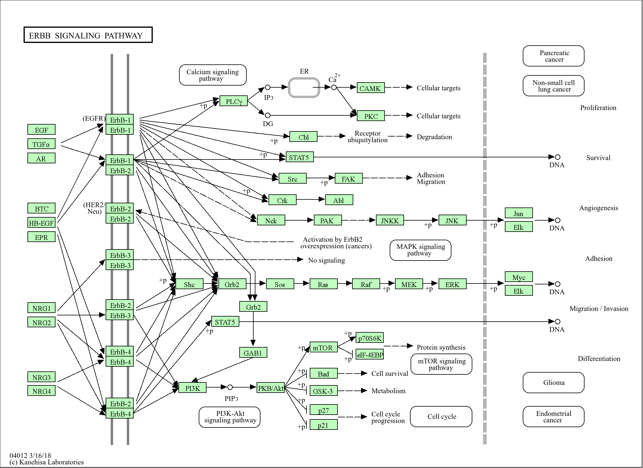
>>NOD-like receptor signaling pathway
show all
Reference Citation({{totalcount}})
Catalog: YP1253
Size
Price
Status
Qty.
200μL
$600.00
In stock
0
100μL
$340.00
In stock
0
50μL
$190.00
In stock
0
Add to cart
Collected
Collect
Recently Viewed Products
Clear allPRODUCTS
CUSTOMIZED
ABOUT US


Toggle night Mode
{{pinfoXq.title || ''}}
Catalog: {{pinfoXq.catalog || ''}}
Filter:
All
{{item.name}}
{{pinfo.title}}
-{{pinfo.catalog}}
Main Information
Target
{{pinfo.target}}
Reactivity
{{pinfo.react}}
Applications
{{pinfo.applicat}}
Conjugate/Modification
{{pinfo.coupling}}/{{pinfo.modific}}
MW (kDa)
{{pinfo.mwcalc}}
Host Species
{{pinfo.hostspec}}
Isotype
{{pinfo.isotype}}
Product {{index}}/{{pcount}}
Prev
Next
{{pvTitle}}
Scroll wheel zooms the picture
{{pvDescr}}