Tropomyosin α Polyclonal Antibody
- Catalog No.:YT6089
- Applications:WB;ELISA
- Reactivity:Human;Mouse;Rat
- Target:
- Tropomyosin α
- Fields:
- >>Cardiac muscle contraction;>>Adrenergic signaling in cardiomyocytes;>>MicroRNAs in cancer;>>Hypertrophic cardiomyopathy;>>Dilated cardiomyopathy
- Gene Name:
- TPM1
- Protein Name:
- Tropomyosin α
- Human Gene Id:
- 7168
- Human Swiss Prot No:
- P09493
- Mouse Gene Id:
- 22003
- Mouse Swiss Prot No:
- P58771
- Immunogen:
- Synthesized peptide derived from human Tropomyosin α. at AA range: 101-150
- Specificity:
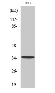
- Tropomyosin α Polyclonal Antibody detects endogenous levels of Tropomyosin α
- Formulation:
- Liquid in PBS containing 50% glycerol, 0.5% BSA and 0.02% sodium azide.
- Source:
- Polyclonal, Rabbit,IgG
- Dilution:
- WB 1:500-2000, ELISA 1:10000-20000
- Purification:
- The antibody was affinity-purified from rabbit antiserum by affinity-chromatography using epitope-specific immunogen.
- Concentration:
- 1 mg/ml
- Storage Stability:
- -15°C to -25°C/1 year(Do not lower than -25°C)
- Other Name:
- Tropomyosin alpha-1 chain (Alpha-tropomyosin) (Tropomyosin-1)
- Observed Band(KD):
- 38kD
- Background:
- This gene is a member of the tropomyosin family of highly conserved, widely distributed actin-binding proteins involved in the contractile system of striated and smooth muscles and the cytoskeleton of non-muscle cells. Tropomyosin is composed of two alpha-helical chains arranged as a coiled-coil. It is polymerized end to end along the two grooves of actin filaments and provides stability to the filaments. The encoded protein is one type of alpha helical chain that forms the predominant tropomyosin of striated muscle, where it also functions in association with the troponin complex to regulate the calcium-dependent interaction of actin and myosin during muscle contraction. In smooth muscle and non-muscle cells, alternatively spliced transcript variants encoding a range of isoforms have been described. Mutations in this gene are associated with type 3 familial hypertrophic cardiomyopathy. [provided by
- Function:
- alternative products:Additional isoforms seem to exist,disease:Defects in TPM1 are the cause of cardiomyopathy dilated type 1Y (CMD1Y) [MIM:611878]. Dilated cardiomyopathy is a disorder characterized by ventricular dilation and impaired systolic function, resulting in congestive heart failure and arrhythmia. Patients are at risk of premature death.,disease:Defects in TPM1 are the cause of cardiomyopathy familial hypertrophic type 3 (CMH3) [MIM:115196]. Familial hypertrophic cardiomyopathy is a hereditary heart disorder characterized by ventricular hypertrophy, which is usually asymmetric and often involves the interventricular septum. The symptoms include dyspnea, syncope, collapse, palpitations, and chest pain. They can be readily provoked by exercise. The disorder has inter- and intrafamilial variability ranging from benign to malignant forms with high risk of cardiac failure and sudde
- Subcellular Location:
- Cytoplasm, cytoskeleton . Associates with F-actin stress fibers. .
- Expression:
- Detected in primary breast cancer tissues but undetectable in normal breast tissues in Sudanese patients. Isoform 1 is expressed in adult and fetal skeletal muscle and cardiac tissues, with higher expression levels in the cardiac tissues. Isoform 10 is expressed in adult and fetal cardiac tissues, but not in skeletal muscle.
- June 19-2018
- WESTERN IMMUNOBLOTTING PROTOCOL
- June 19-2018
- IMMUNOHISTOCHEMISTRY-PARAFFIN PROTOCOL
- June 19-2018
- IMMUNOFLUORESCENCE PROTOCOL
- September 08-2020
- FLOW-CYTOMEYRT-PROTOCOL
- May 20-2022
- Cell-Based ELISA│解您多样本WB检测之困扰
- July 13-2018
- CELL-BASED-ELISA-PROTOCOL-FOR-ACETYL-PROTEIN
- July 13-2018
- CELL-BASED-ELISA-PROTOCOL-FOR-PHOSPHO-PROTEIN
- July 13-2018
- Antibody-FAQs
- Products Images
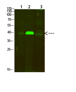
- Western Blot analysis of 1,mouse-lung 2,mouse-brain 3,mouse-spleen cells using primary antibody diluted at 1:500(4°C overnight). Secondary antibody:Goat Anti-rabbit IgG IRDye 800( diluted at 1:5000, 25°C, 1 hour)