CD81 Polyclonal Antibody
- Catalog No.:YT5394
- Applications:IF;WB;ELISA
- Reactivity:Human;Mouse;Rat
- Target:
- CD81
- Fields:
- >>B cell receptor signaling pathway;>>Malaria;>>Hepatitis C
- Gene Name:
- CD81
- Protein Name:
- CD81 antigen
- Human Gene Id:
- 975
- Human Swiss Prot No:
- P60033
- Mouse Gene Id:
- 12520
- Mouse Swiss Prot No:
- P35762
- Rat Gene Id:
- 25621
- Rat Swiss Prot No:
- Q62745
- Immunogen:
- The antiserum was produced against synthesized peptide derived from the Internal region of human CD81. AA range:111-160
- Specificity:
- CD81 Polyclonal Antibody detects endogenous levels of CD81 protein.
- Formulation:
- Liquid in PBS containing 50% glycerol, 0.5% BSA and 0.02% sodium azide.
- Source:
- Polyclonal, Rabbit,IgG
- Dilution:
- IF 1:50-200 WB 1:500 - 1:2000. ELISA: 1:20000. Not yet tested in other applications.
- Purification:
- The antibody was affinity-purified from rabbit antiserum by affinity-chromatography using epitope-specific immunogen.
- Concentration:
- 1 mg/ml
- Storage Stability:
- -15°C to -25°C/1 year(Do not lower than -25°C)
- Other Name:
- CD81;TAPA1;TSPAN28;CD81 antigen;26 kDa cell surface protein TAPA-1;Target of the antiproliferative antibody 1;Tetraspanin-28;Tspan-28;CD81
- Observed Band(KD):
- 26kD
- Background:
- The protein encoded by this gene is a member of the transmembrane 4 superfamily, also known as the tetraspanin family. Most of these members are cell-surface proteins that are characterized by the presence of four hydrophobic domains. The proteins mediate signal transduction events that play a role in the regulation of cell development, activation, growth and motility. This encoded protein is a cell surface glycoprotein that is known to complex with integrins. This protein appears to promote muscle cell fusion and support myotube maintenance. Also it may be involved in signal transduction. This gene is localized in the tumor-suppressor gene region and thus it is a candidate gene for malignancies. Two transcript variants encoding different isoforms have been found for this gene. [provided by RefSeq, Jul 2014],
- Function:
- function:May play an important role in the regulation of lymphoma cell growth. Interacts with a 16-kDa Leu-13 protein to form a complex possibly involved in signal transduction. May acts a the viral receptor for HCV.,PTM:Not glycosylated.,similarity:Belongs to the tetraspanin (TM4SF) family.,subunit:Plays a critical role in HCV attachment and/or cell entry by interacting with HCV E1/E2 glycoproteins heterodimer. Interacts directly with IGSF8.,tissue specificity:Hematolymphoid, neuroectodermal and mesenchymal tumor cell lines.,
- Subcellular Location:
- Cell membrane ; Multi-pass membrane protein . Basolateral cell membrane ; Multi-pass membrane protein . Associates with CLDN1 and the CLDN1-CD81 complex localizes to the basolateral cell membrane. .
- Expression:
- Expressed on B cells (at protein level) (PubMed:20237408). Expressed in hepatocytes (at protein level) (PubMed:12483205). Expressed in monocytes/macrophages (at protein level) (PubMed:12796480). Expressed on both naive and memory CD4-positive T cells (at protein level) (PubMed:22307619).
The preliminary study of exosomes derived from thymosin beta 4-treated adipose-derived stem cells in fat grafting
Exosomal transfer of miR-181b-5p confers senescence-mediated doxorubicin resistance via modulating BCLAF1 in breast cancer BRITISH JOURNAL OF CANCER Zhao, Shaorong, Pan, Teng, Deng, Jinhai, Cao, Lixia, Vicencio, Jose M., Liu, Jingjing, Zhou, Guanglin, Ng, Tony, Zhang, Jin WB Human Cal51 cell, MCF-7 cell
Serum exosomal proteomics analysis of lung adenocarcinoma to discover new tumor markers Bmc Cancer. 2022 Dec;22(1):1-11. WB Human 1:1000 serum exosomal
Adipose-derived mesenchymal stem cells and extracellular vesicles confer antitumor activity in preclinical treatment of breast cancer. PHARMACOLOGICAL RESEARCH 2020 Apr 30 WB Mouse ADSCs, CD90high ADSC‑EVs,CD90low ADSC‑EVs
Mesenchymal stem cell‑derived extracellular vesicles promote the in vitro proliferation and migration of breast cancer cells through the activation of the ERK pathway. INTERNATIONAL JOURNAL OF ONCOLOGY Int J Oncol. 2019 May;54(5):1843-1852 WB Human 1:1000 hUC-MSC-EVs
CD44-targeting Drug Delivery System of Exosomes Loading Forsythiaside A Combats Liver Fibrosis via Regulating NLRP3-mediated Pyroptosis Advanced Healthcare Materials Cheng Peng WB Bovine exosomes
Phillygenin inhibited M1 macrophage polarization and reduced hepatic stellate cell activation by inhibiting macrophage exosomal miR-125b-5p BIOMEDICINE & PHARMACOTHERAPY Cheng Ma WB Mouse RAW264.7 cells
Exosomal transfer leads to chemoresistance through oxidative phosphorylation-mediated stemness phenotype in colorectal cancer. Theranostics Mingzhu Yin WB Human 1:1000 HCT15 cell,DLD1 cell
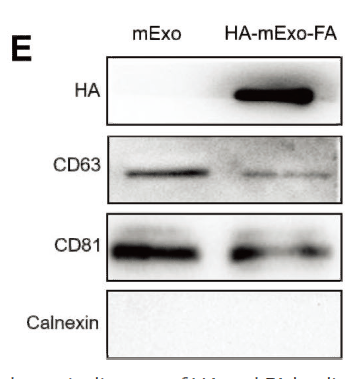
Preparation of Phillygenin-Hyaluronic acid composite milk-derived exosomes and its anti-hepatic fibrosis effect. Materials Today Bio Yunxia Li WB Bovine 1:1000 Milk-derived exosomes (mEXO)
The Influence of Extracellular Vesicles Secreted by Dural Cells on Osteoblasts MOLECULAR BIOTECHNOLOGY Zhao Fangning WB Mouse extracellular vesicles secreted by dural cells (Dura-EVs)
- June 19-2018
- WESTERN IMMUNOBLOTTING PROTOCOL
- June 19-2018
- IMMUNOHISTOCHEMISTRY-PARAFFIN PROTOCOL
- June 19-2018
- IMMUNOFLUORESCENCE PROTOCOL
- September 08-2020
- FLOW-CYTOMEYRT-PROTOCOL
- May 20-2022
- Cell-Based ELISA│解您多样本WB检测之困扰
- July 13-2018
- CELL-BASED-ELISA-PROTOCOL-FOR-ACETYL-PROTEIN
- July 13-2018
- CELL-BASED-ELISA-PROTOCOL-FOR-PHOSPHO-PROTEIN
- July 13-2018
- Antibody-FAQs
- Products Images
- CD44-targeting Drug Delivery System of Exosomes Loading Forsythiaside A Combats Liver Fibrosis via Regulating NLRP3-mediated Pyroptosis Advanced Healthcare Materials Cheng Peng WB Bovine exosomes
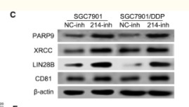
- Wang, Xinyi, et al. "Exosomes serve as nanoparticles to deliver anti-miR-214 to reverse chemoresistance to cisplatin in gastric cancer." Molecular Therapy 26.3 (2018): 774-783.
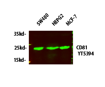
- Western blot analysis of lysates from HT-29, NIH/3T3, and HepG2 cells, primary antibody was diluted at 1:1000, 4° over night, secondary antibody(cat: RS23920)was diluted at 1:10000, 37° 1hour.
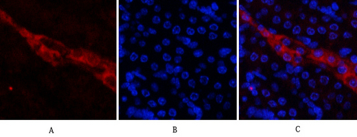
- Immunofluorescence analysis of mouse-kidney tissue. 1,CD81 Polyclonal Antibody(red) was diluted at 1:200(4°C,overnight). 2, Cy3 labled Secondary antibody was diluted at 1:300(room temperature, 50min).3, Picture B: DAPI(blue) 10min. Picture A:Target. Picture B: DAPI. Picture C: merge of A+B
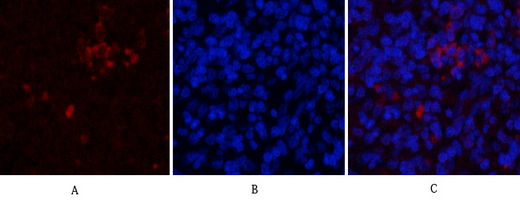
- Immunofluorescence analysis of rat-spleen tissue. 1,CD81 Polyclonal Antibody(red) was diluted at 1:200(4°C,overnight). 2, Cy3 labled Secondary antibody was diluted at 1:300(room temperature, 50min).3, Picture B: DAPI(blue) 10min. Picture A:Target. Picture B: DAPI. Picture C: merge of A+B
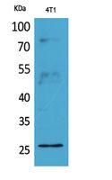
- Western Blot analysis of 4T1 cells using CD81 Polyclonal Antibody. Antibody was diluted at 1:2000. Secondary antibody(catalog#:RS0002) was diluted at 1:20000
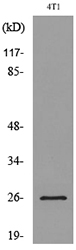
- Western blot analysis of lysate from 4T1 cells, using CD81 Antibody.


