Stathmin-2 Polyclonal Antibody
- Catalog No.:YT5076
- Applications:WB;IHC;IF;ELISA
- Reactivity:Human;Mouse;Rat
- Target:
- Stathmin-2
- Gene Name:
- STMN2
- Protein Name:
- Stathmin-2
- Human Gene Id:
- 11075
- Human Swiss Prot No:
- Q93045
- Mouse Gene Id:
- 20257
- Mouse Swiss Prot No:
- P55821
- Rat Gene Id:
- 84510
- Rat Swiss Prot No:
- P21818
- Immunogen:
- Synthesized peptide derived from the Internal region of human Stathmin-2.
- Specificity:
- Stathmin-2 Polyclonal Antibody detects endogenous levels of Stathmin-2 protein.
- Formulation:
- Liquid in PBS containing 50% glycerol, 0.5% BSA and 0.02% sodium azide.
- Source:
- Polyclonal, Rabbit,IgG
- Dilution:
- WB 1:500 - 1:2000. IHC: 1:100-300 ELISA: 1:5000.. IF 1:50-200
- Purification:
- The antibody was affinity-purified from rabbit antiserum by affinity-chromatography using epitope-specific immunogen.
- Concentration:
- 1 mg/ml
- Storage Stability:
- -15°C to -25°C/1 year(Do not lower than -25°C)
- Other Name:
- STMN2;SCG10;SCGN10;Stathmin-2;Superior cervical ganglion-10 protein;Protein SCG10
- Observed Band(KD):
- 20kD
- Background:
- This gene encodes a member of the stathmin family of phosphoproteins. Stathmin proteins function in microtubule dynamics and signal transduction. The encoded protein plays a regulatory role in neuronal growth and is also thought to be involved in osteogenesis. Reductions in the expression of this gene have been associated with Down's syndrome and Alzheimer's disease. Alternatively spliced transcript variants have been observed for this gene. A pseudogene of this gene is located on the long arm of chromosome 6. [provided by RefSeq, Nov 2010],
- Function:
- function:May play a role in neuronal differentiation, and in modulating membrane interaction with the cytoskeleton during neurite outgrowth.,PTM:Sumoylated.,similarity:Belongs to the stathmin family.,subcellular location:Associated with punctate structures in the perinuclear cytoplasm, axons, and growth cones of developing neurons. SCG10 exists in both soluble and membrane-bound forms.,tissue specificity:Neuron specific.,
- Subcellular Location:
- Cytoplasm . Cytoplasm, perinuclear region . Cell projection, growth cone. Membrane ; Peripheral membrane protein ; Cytoplasmic side . Cell projection, axon. Golgi apparatus. Endosome . Cell projection, lamellipodium. Associated with punctate structures in the perinuclear cytoplasm, axons, and growth cones of developing neurons. SCG10 exists in both soluble and membrane-bound forms. Colocalized with CIB1 in neurites of developing hippocampal primary neurons (By similarity). Colocalized with CIB1 in the cell body, neuritis and growth cones of neurons. Colocalized with CIB1 to the leading edge of lamellipodia. .
- Expression:
- Neuron specific.
- June 19-2018
- WESTERN IMMUNOBLOTTING PROTOCOL
- June 19-2018
- IMMUNOHISTOCHEMISTRY-PARAFFIN PROTOCOL
- June 19-2018
- IMMUNOFLUORESCENCE PROTOCOL
- September 08-2020
- FLOW-CYTOMEYRT-PROTOCOL
- May 20-2022
- Cell-Based ELISA│解您多样本WB检测之困扰
- July 13-2018
- CELL-BASED-ELISA-PROTOCOL-FOR-ACETYL-PROTEIN
- July 13-2018
- CELL-BASED-ELISA-PROTOCOL-FOR-PHOSPHO-PROTEIN
- July 13-2018
- Antibody-FAQs
- Products Images

- Western Blot analysis of extracts from Jurkat cells, using Stathmin-2 Polyclonal Antibody. Secondary antibody(catalog#:RS0002) was diluted at 1:20000
.jpg)
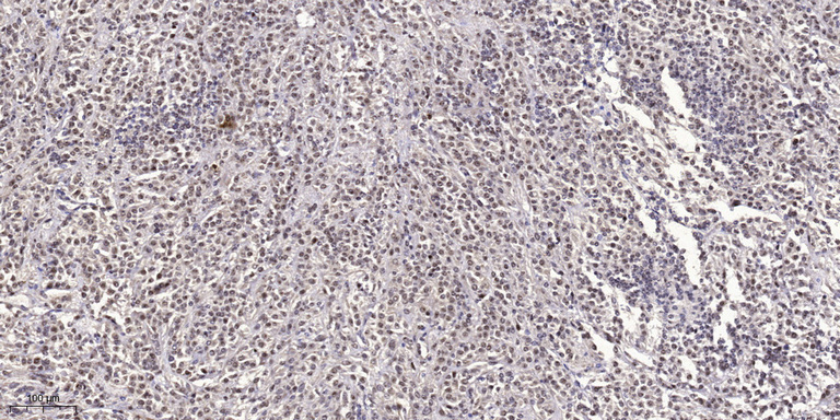
- Immunohistochemical analysis of paraffin-embedded rat-brain, antibody was diluted at 1:100
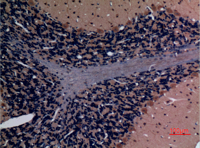
- Immunohistochemical analysis of paraffin-embedded rat-brain, antibody was diluted at 1:100