Di-Ras1 Polyclonal Antibody
- Catalog No.:YT1353
- Applications:WB;IHC;IF;ELISA
- Reactivity:Human;Rat;Mouse;
- Target:
- Di-Ras1
- Gene Name:
- DIRAS1
- Protein Name:
- GTP-binding protein Di-Ras1
- Human Gene Id:
- 148252
- Human Swiss Prot No:
- O95057
- Mouse Swiss Prot No:
- Q91Z61
- Immunogen:
- The antiserum was produced against synthesized peptide derived from human DIRA1. AA range:149-198
- Specificity:
- Di-Ras1 Polyclonal Antibody detects endogenous levels of Di-Ras1 protein.
- Formulation:
- Liquid in PBS containing 50% glycerol, 0.5% BSA and 0.02% sodium azide.
- Source:
- Polyclonal, Rabbit,IgG
- Dilution:
- WB 1:500 - 1:2000. IHC 1:100 - 1:300. IF 1:200 - 1:1000. ELISA: 1:20000. Not yet tested in other applications.
- Purification:
- The antibody was affinity-purified from rabbit antiserum by affinity-chromatography using epitope-specific immunogen.
- Concentration:
- 1 mg/ml
- Storage Stability:
- -15°C to -25°C/1 year(Do not lower than -25°C)
- Other Name:
- DIRAS1;GBTS1;RIG;GTP-binding protein Di-Ras1;Distinct subgroup of the Ras family member 1;Ras-related inhibitor of cell growth;Rig;Small GTP-binding tumor suppressor 1
- Observed Band(KD):
- 22kD
- Background:
- DIRAS1 belongs to a distinct branch of the functionally diverse Ras (see HRAS; MIM 190020) superfamily of monomeric GTPases.[supplied by OMIM, Apr 2004],
- Function:
- function:Displays low GTPase activity and exist predominantly in the GTP-bound form.,similarity:Belongs to the small GTPase superfamily. Di-Ras family.,tissue specificity:Highly expressed in heart and brain.,
- Subcellular Location:
- Cell membrane ; Lipid-anchor ; Cytoplasmic side .
- Expression:
- Highly expressed in heart and brain.
- June 19-2018
- WESTERN IMMUNOBLOTTING PROTOCOL
- June 19-2018
- IMMUNOHISTOCHEMISTRY-PARAFFIN PROTOCOL
- June 19-2018
- IMMUNOFLUORESCENCE PROTOCOL
- September 08-2020
- FLOW-CYTOMEYRT-PROTOCOL
- May 20-2022
- Cell-Based ELISA│解您多样本WB检测之困扰
- July 13-2018
- CELL-BASED-ELISA-PROTOCOL-FOR-ACETYL-PROTEIN
- July 13-2018
- CELL-BASED-ELISA-PROTOCOL-FOR-PHOSPHO-PROTEIN
- July 13-2018
- Antibody-FAQs
- Products Images
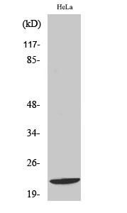
- Western Blot analysis of various cells using Di-Ras1 Polyclonal Antibody
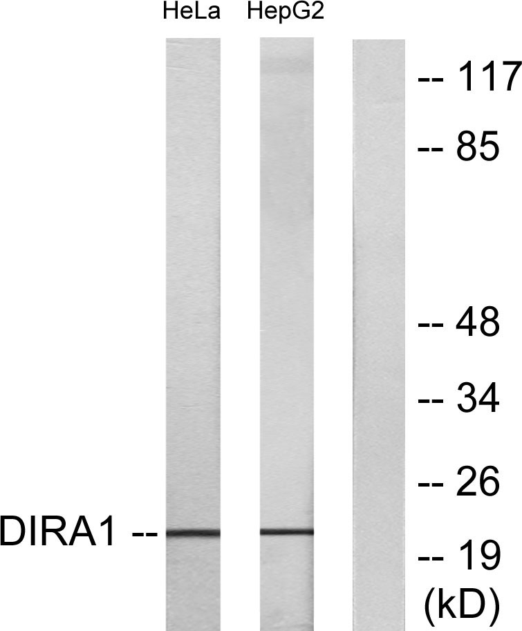
- Western blot analysis of lysates from HeLa and HepG2 cells, using DIRA1 Antibody. The lane on the right is blocked with the synthesized peptide.
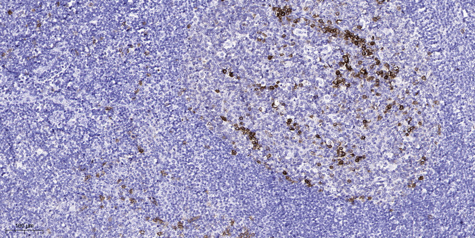
- Immunohistochemical analysis of paraffin-embedded human tonsil. 1, Antibody was diluted at 1:200(4° overnight). 2, Tris-EDTA,pH9.0 was used for antigen retrieval. 3,Secondary antibody was diluted at 1:200(room temperature, 45min).


