17β-HSD11 Polyclonal Antibody
- Catalog No.:YT0013
- Applications:WB;IF;ELISA
- Reactivity:Human;Rat;Mouse;
- Target:
- 17β-HSD11
- Gene Name:
- HSD17B11
- Protein Name:
- Estradiol 17-beta-dehydrogenase 11
- Human Gene Id:
- 51170
- Human Swiss Prot No:
- Q8NBQ5
- Mouse Swiss Prot No:
- Q9EQ06
- Immunogen:
- The antiserum was produced against synthesized peptide derived from human DHRS8. AA range:71-120
- Specificity:
- 17β-HSD11 Polyclonal Antibody detects endogenous levels of 17β-HSD11 protein.
- Formulation:
- Liquid in PBS containing 50% glycerol, 0.5% BSA and 0.02% sodium azide.
- Source:
- Polyclonal, Rabbit,IgG
- Dilution:
- WB 1:500 - 1:2000. ELISA: 1:5000. IF 1:100-300 Not yet tested in other applications.
- Purification:
- The antibody was affinity-purified from rabbit antiserum by affinity-chromatography using epitope-specific immunogen.
- Concentration:
- 1 mg/ml
- Storage Stability:
- -15°C to -25°C/1 year(Do not lower than -25°C)
- Other Name:
- HSD17B11;DHRS8;PAN1B;PSEC0029;Estradiol 17-beta-dehydrogenase 11;17-beta-hydroxysteroid dehydrogenase 11;17-beta-HSD 11;17bHSD11;17betaHSD11;17-beta-hydroxysteroid dehydrogenase XI;17-beta-HSD XI;17betaHSDXI;Cutaneous T-cell lym
- Observed Band(KD):
- 36kD
- Background:
- Short-chain alcohol dehydrogenases, such as HSD17B11, metabolize secondary alcohols and ketones (Brereton et al., 2001 [PubMed 11165019]).[supplied by OMIM, Jun 2009],
- Function:
- catalytic activity:Estradiol-17-beta + NAD(P)(+) = estrone + NAD(P)H.,function:Can convert androstan-3-alpha,17-beta-diol (3-alpha-diol) to androsterone in vitro, suggesting that it may participate in androgen metabolism during steroidogenesis. May act by metabolizing compounds that stimulate steroid synthesis and/or by generating metabolites that inhibit it. Has no activity toward DHEA (dehydroepiandrosterone), or A-dione (4-androste-3,17-dione), and only a slight activity toward testosterone to A-dione. Tumor-associated antigen in cutaneous T-cell lymphoma.,similarity:Belongs to the short-chain dehydrogenases/reductases (SDR) family. 17-beta-HSD 3 subfamily.,tissue specificity:Present at high level in steroidogenic cells such as syncytiotrophoblasts, sebaceous gland, Leydig cells, and granulosa cells of the dominant follicle and corpus luteum. In lung, it is detected in the ciliated ep
- Subcellular Location:
- Endoplasmic reticulum . Lipid droplet . Redistributed from the endoplasmic reticulum to lipids droplets in the cell upon induction of lipids droplet formation. .
- Expression:
- Present at high level in steroidogenic cells such as syncytiotrophoblasts, sebaceous gland, Leydig cells, and granulosa cells of the dominant follicle and corpus luteum. In lung, it is detected in the ciliated epithelium and in acini of adult trachea, in bronchioles, but not in alveoli. In the eye, it is detected in the nonpigmented epithelium of the ciliary body and, at lower level, in the inner nuclear layer of the retina (at protein level). Widely expressed. Highly expressed in retina, pancreas, kidney, liver, lung, adrenal, small intestine, ovary and heart.
Alleviating effects of selenium on fluoride-induced testosterone synthesis disorder and reproduction toxicity in rats
MicroRNA‑143 increases cell apoptosis in myelodysplastic syndrome through the Fas/FasL pathway both in vitro and in vivo. INTERNATIONAL JOURNAL OF ONCOLOGY Int J Oncol. 2018 Nov;53(5):2191-2199 WB Human SKM-1 cell
- June 19-2018
- WESTERN IMMUNOBLOTTING PROTOCOL
- June 19-2018
- IMMUNOHISTOCHEMISTRY-PARAFFIN PROTOCOL
- June 19-2018
- IMMUNOFLUORESCENCE PROTOCOL
- September 08-2020
- FLOW-CYTOMEYRT-PROTOCOL
- May 20-2022
- Cell-Based ELISA│解您多样本WB检测之困扰
- July 13-2018
- CELL-BASED-ELISA-PROTOCOL-FOR-ACETYL-PROTEIN
- July 13-2018
- CELL-BASED-ELISA-PROTOCOL-FOR-PHOSPHO-PROTEIN
- July 13-2018
- Antibody-FAQs
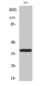
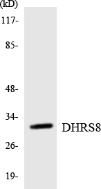
- Products Images
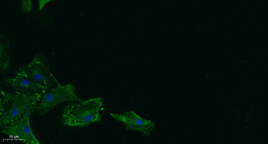
- Immunofluorescence analysis of A549. 1,primary Antibody was diluted at 1:200(4°C overnight). 2, Goat Anti Rabbit IgG (H&L) - Alexa Fluor 488 Secondary antibody was diluted at 1:1000(room temperature, 50min).3, Picture B: DAPI(blue) 10min.
- Western Blot analysis of various cells using 17β-HSD11 Polyclonal Antibody

- Western blot analysis of lysates from 293 cells, using DHRS8 Antibody. The lane on the right is blocked with the synthesized peptide.
- Western blot analysis of the lysates from RAW264.7cells using DHRS8 antibody.


