20S Proteasome α3 (phospho Ser250) Polyclonal Antibody
- Catalog No.:YP0396
- Applications:WB;IHC;IF;ELISA
- Reactivity:Human;Mouse;Rat
- Target:
- Proteasome α3
- Fields:
- >>Proteasome;>>Alzheimer disease;>>Parkinson disease;>>Amyotrophic lateral sclerosis;>>Huntington disease;>>Spinocerebellar ataxia;>>Prion disease;>>Pathways of neurodegeneration - multiple diseases
- Gene Name:
- PSMA3
- Protein Name:
- Proteasome subunit alpha type-3
- Human Gene Id:
- 5684
- Human Swiss Prot No:
- P25788
- Mouse Gene Id:
- 19167
- Mouse Swiss Prot No:
- O70435
- Rat Gene Id:
- 29670
- Rat Swiss Prot No:
- P18422
- Immunogen:
- The antiserum was produced against synthesized peptide derived from human Proteasome alpha3 around the phosphorylation site of Ser250. AA range:206-255
- Specificity:
- Phospho-20S Proteasome α3 (S250) Polyclonal Antibody detects endogenous levels of 20S Proteasome α3 protein only when phosphorylated at S250.
- Formulation:
- Liquid in PBS containing 50% glycerol, 0.5% BSA and 0.02% sodium azide.
- Source:
- Polyclonal, Rabbit,IgG
- Dilution:
- WB 1:500 - 1:2000. IHC 1:100 - 1:300. ELISA: 1:10000.. IF 1:50-200
- Purification:
- The antibody was affinity-purified from rabbit antiserum by affinity-chromatography using epitope-specific immunogen.
- Concentration:
- 1 mg/ml
- Storage Stability:
- -15°C to -25°C/1 year(Do not lower than -25°C)
- Other Name:
- PSMA3;HC8;PSC8;Proteasome subunit alpha type-3;Macropain subunit C8;Multicatalytic endopeptidase complex subunit C8;Proteasome component C8
- Observed Band(KD):
- 32kD
- Background:
- The proteasome is a multicatalytic proteinase complex with a highly ordered ring-shaped 20S core structure. The core structure is composed of 4 rings of 28 non-identical subunits; 2 rings are composed of 7 alpha subunits and 2 rings are composed of 7 beta subunits. Proteasomes are distributed throughout eukaryotic cells at a high concentration and cleave peptides in an ATP/ubiquitin-dependent process in a non-lysosomal pathway. An essential function of a modified proteasome, the immunoproteasome, is the processing of class I MHC peptides. This gene encodes a member of the peptidase T1A family, that is a 20S core alpha subunit. Two alternative transcripts encoding different isoforms have been identified. [provided by RefSeq, Jul 2008],
- Function:
- catalytic activity:Cleavage of peptide bonds with very broad specificity.,function:The proteasome is a multicatalytic proteinase complex which is characterized by its ability to cleave peptides with Arg, Phe, Tyr, Leu, and Glu adjacent to the leaving group at neutral or slightly basic pH. The proteasome has an ATP-dependent proteolytic activity.,similarity:Belongs to the peptidase T1A family.,subunit:The 26S proteasome consists of a 20S proteasome core and two 19S regulatory subunits. The 20S proteasome core is composed of 28 subunits that are arranged in four stacked rings, resulting in a barrel-shaped structure. The two end rings are each formed by seven alpha subunits, and the two central rings are each formed by seven beta subunits.,subunit:The 26S proteasome consists of a 20S proteasome core and two 19S regulatory subunits. The 20S proteasome core is composed of 28 subunits that are
- Subcellular Location:
- Cytoplasm . Nucleus . Translocated from the cytoplasm into the nucleus following interaction with AKIRIN2, which bridges the proteasome with the nuclear import receptor IPO9. .
- Expression:
- Bone marrow,Epithelium,Liver,Pancreas,Urinary bladder,
- June 19-2018
- WESTERN IMMUNOBLOTTING PROTOCOL
- June 19-2018
- IMMUNOHISTOCHEMISTRY-PARAFFIN PROTOCOL
- June 19-2018
- IMMUNOFLUORESCENCE PROTOCOL
- September 08-2020
- FLOW-CYTOMEYRT-PROTOCOL
- May 20-2022
- Cell-Based ELISA│解您多样本WB检测之困扰
- July 13-2018
- CELL-BASED-ELISA-PROTOCOL-FOR-ACETYL-PROTEIN
- July 13-2018
- CELL-BASED-ELISA-PROTOCOL-FOR-PHOSPHO-PROTEIN
- July 13-2018
- Antibody-FAQs
- Products Images
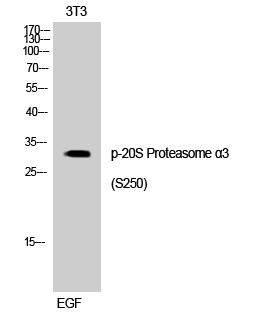
- Western Blot analysis of 3T3 cells using Phospho-20S Proteasome α3 (S250) Polyclonal Antibody
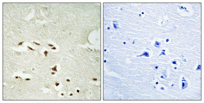
- Immunohistochemical analysis of paraffin-embedded Human brain. Antibody was diluted at 1:100(4° overnight). High-pressure and temperature Tris-EDTA,pH8.0 was used for antigen retrieval. Negetive contrl (right) obtaned from antibody was pre-absorbed by immunogen peptide.

- Western blot analysis of lysates from NIH/3T3 cells treated with EGF 200ng/ml 30', using Proteasome alpha3 (Phospho-Ser250) Antibody. The lane on the right is blocked with the phospho peptide.


