IκB-α (phospho Ser32/S36) Polyclonal Antibody
- Catalog No.:YP0151
- Applications:WB;IHC;IF;ELISA
- Reactivity:Human;Mouse;Rat;Monkey
- Target:
- IκB-α
- Fields:
- >>cAMP signaling pathway;>>Chemokine signaling pathway;>>NF-kappa B signaling pathway;>>Apoptosis;>>Osteoclast differentiation;>>Toll-like receptor signaling pathway;>>NOD-like receptor signaling pathway;>>RIG-I-like receptor signaling pathway;>>Cytosolic DNA-sensing pathway;>>C-type lectin receptor signaling pathway;>>IL-17 signaling pathway;>>Th1 and Th2 cell differentiation;>>Th17 cell differentiation;>>T cell receptor signaling pathway;>>B cell receptor signaling pathway;>>TNF signaling pathway;>>Neurotrophin signaling pathway;>>Adipocytokine signaling pathway;>>Relaxin signaling pathway;>>Insulin resistance;>>Alcoholic liver disease;>>Epithelial cell signaling in Helicobacter pylori infection;>>Pathogenic Escherichia coli infection;>>Shigellosis;>>Salmonella infection;>>Legionellosis;>>Yersinia infection;>>Leishmaniasis;>>Chagas disease;>>Toxoplasmosis;>>Hepatitis C;>>Hepatitis B;>>Measles;>>Human cytomegalovirus infection;>>Influenza A;>>Human T-cell leukemia virus 1 infection;>>
- Gene Name:
- NFKBIA IKBA MAD3 NFKBI
- Protein Name:
- NF-kappa-B inhibitor alpha
- Human Gene Id:
- 4792
- Human Swiss Prot No:
- P25963
- Mouse Gene Id:
- 18035
- Mouse Swiss Prot No:
- Q9Z1E3
- Rat Gene Id:
- 25493
- Rat Swiss Prot No:
- Q63746
- Immunogen:
- The antiserum was produced against synthesized peptide derived from human IkappaB-alpha around the phosphorylation site of Ser32/Ser36. AA range:15-64
- Specificity:
- Phospho-IκB-α (S32/S36) Polyclonal Antibody detects endogenous levels of IκB-α protein only when phosphorylated at S32/S36.
- Formulation:
- Liquid in PBS containing 50% glycerol, 0.5% BSA and 0.02% sodium azide.
- Source:
- Polyclonal, Rabbit,IgG
- Dilution:
- WB 1:500 - 1:2000. IHC 1:100 - 1:300. IF 1:200 - 1:1000. ELISA: 1:10000. Not yet tested in other applications.
- Purification:
- The antibody was affinity-purified from rabbit antiserum by affinity-chromatography using epitope-specific immunogen.
- Concentration:
- 1 mg/ml
- Storage Stability:
- -15°C to -25°C/1 year(Do not lower than -25°C)
- Other Name:
- NFKBIA;IKBA;MAD3;NFKBI;NF-kappa-B inhibitor alpha;I-kappa-B-alpha;IkB-alpha;IkappaBalpha;Major histocompatibility complex enhancer-binding protein MAD3
- Observed Band(KD):
- about 40kd
- Background:
- This gene encodes a member of the NF-kappa-B inhibitor family, which contain multiple ankrin repeat domains. The encoded protein interacts with REL dimers to inhibit NF-kappa-B/REL complexes which are involved in inflammatory responses. The encoded protein moves between the cytoplasm and the nucleus via a nuclear localization signal and CRM1-mediated nuclear export. Mutations in this gene have been found in ectodermal dysplasia anhidrotic with T-cell immunodeficiency autosomal dominant disease. [provided by RefSeq, Aug 2011],
- Function:
- disease:Defects in NFKBIA are the cause of ectodermal dysplasia anhidrotic with T-cell immunodeficiency autosomal dominant (ADEDAID) [MIM:612132]. Ectodermal dysplasia defines a heterogeneous group of disorders due to abnormal development of two or more ectodermal structures. ADEDAID is an ectodermal dysplasia associated with decreased production of pro-inflammatory cytokines and certain interferons, rendering patients susceptible to infection.,function:Inhibits the activity of dimeric NF-kappa-B/REL complexes by trapping REL dimers in the cytoplasm through masking of their nuclear localization signals. On cellular stimulation by immune and proinflammatory responses, becomes phosphorylated promoting ubiquitination and degradation, enabling the dimeric RELA to tranlocate to the nucleus and activate transcription.,induction:Induced in adherent monocytes.,online information:NFKBIA mutation
- Subcellular Location:
- Cytoplasm. Nucleus. Shuttles between the nucleus and the cytoplasm by a nuclear localization signal (NLS) and a CRM1-dependent nuclear export. .
- Expression:
- Brain,Kidney,Lymph node,Monocyte,
Tyrosol inhibits NF-κB pathway in the treatment of enterotoxigenic Escherichia coli-induced diarrhea in mice MICROBIAL PATHOGENESIS Fazheng Yu, Jian Guo, Hong lin Ren, Shiying Lu, Zhaoqi He, Jiang Chang, Xueyu Hu, Ruoran Shi, Yuanyuan Jin, Yansong Li, Zengshan Liu, Xiaoxu Wang, Pan Hu WB Mouse colonic tissue
MAST3 modulates the inflammatory response and proliferation of fibroblast-like synoviocytes in rheumatoid arthritis. INTERNATIONAL IMMUNOPHARMACOLOGY Int Immunopharmacol. 2019 Dec;77:105900 WB Rat 1:500 Synovial tissues FLSs
NLRC5 promotes cell proliferation via regulating the NF-κB signaling pathway in Rheumatoid arthritis. MOLECULAR IMMUNOLOGY Mol Immunol. 2017 Nov;91:24 WB Rat 1:500 Synovial tissues FLS cell
Lactoferrin suppresses lipopolysaccharide-induced endometritis in mice via down-regulation of the NF-κB pathway. INTERNATIONAL IMMUNOPHARMACOLOGY Int Immunopharmacol. 2015 Sep;28:695 WB Mouse 1:1000 uterine tissues
PSTPIP2 Inhibits the Inflammatory Response and Proliferation of Fibroblast-Like Synoviocytes in vitro. Frontiers in Pharmacology Front Pharmacol. 2018 Dec;9:1432 WB Rat 1:1000 Synovial tissues Fibroblast-like synoviocytes (FLSs)
Activation of NLRP3 inflammasome up-regulates TREM-1 expression in murine macrophages via HMGB1 and IL-18. INTERNATIONAL IMMUNOPHARMACOLOGY Int Immunopharmacol. 2020 Dec;89:107045 WB Mouse 1:1000 Lung Macrophage
miR‐377‐3p drives malignancy characteristics via upregulating GSK‐3β expression and activating NF‐κB pathway in hCRC cells. JOURNAL OF CELLULAR BIOCHEMISTRY J Cell Biochem. 2018 Feb;119(2):2124-2134 WB Human HCT116 cell,SW480 cell
Dopamine receptor D2 inhibition alleviates diabetic hepatic stellate cells fibrosis by regulating the TGF‐β1/Smads and NFκB pathways. CLINICAL AND EXPERIMENTAL PHARMACOLOGY AND PHYSIOLOGY Clin Exp Pharmacol P. 2021 Mar;48(3):370-380 WB Rat 1 : 1000 Liver hepatic stellate cells (HSCs)
Liao, Jiamei, et al. "Anti-inflammatory Activity of Essential Oil from Leaves of Blumea balsamifera (L.) DC through Inhibiting TLR4/NF-kB Signaling Pathways and NLRP3 Inflammasome Activation in LPS-induced RAW264. 7 Macrophage Cells." Journal of Essential Oil Bearing Plants (2021): 1-17.
The adhesion protein of Mycoplasma genitalium inhibits urethral epithelial cell apoptosis through CypA-CD147 activating PI3K/ Akt/NF-κB pathway APPLIED MICROBIOLOGY AND BIOTECHNOLOGY Yanhua Zeng WB Human
Surface functionalization-dependent inflammatory potential of polystyrene nanoplastics through the activation of MAPK/ NF-κB signaling pathways in macrophage Raw 264.7 ECOTOXICOLOGY AND ENVIRONMENTAL SAFETY Jian Xu WB Mouse RAW 264.7 cells
Effects of Aire on perforin expression in BMDCs via TLR7/8 and its therapeutic effect on type 1 diabetes INTERNATIONAL IMMUNOPHARMACOLOGY Wei Yang WB Mouse bone marrow-derived dendritic cells (BMDCs)
Oridonin Attenuates Thioacetamide-Induced Osteoclastogenesis Through MAPK/NF-κB Pathway and Thioacetamide-Inhibited Osteoblastogenesis Through BMP-2/RUNX2 Pathway CALCIFIED TISSUE INTERNATIONAL Xu Jian WB Mouse,Rat RAW264.7 cell,bone mesenchymal stem cells (BMSCs)
- June 19-2018
- WESTERN IMMUNOBLOTTING PROTOCOL
- June 19-2018
- IMMUNOHISTOCHEMISTRY-PARAFFIN PROTOCOL
- June 19-2018
- IMMUNOFLUORESCENCE PROTOCOL
- September 08-2020
- FLOW-CYTOMEYRT-PROTOCOL
- May 20-2022
- Cell-Based ELISA│解您多样本WB检测之困扰
- July 13-2018
- CELL-BASED-ELISA-PROTOCOL-FOR-ACETYL-PROTEIN
- July 13-2018
- CELL-BASED-ELISA-PROTOCOL-FOR-PHOSPHO-PROTEIN
- July 13-2018
- Antibody-FAQs
- Products Images
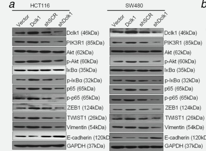
- Liu, Weiying, et al. "DCLK1 promotes epithelial‐mesenchymal transition via the PI3K/Akt/NF‐κB pathway in colorectal cancer." International journal of cancer 142.10 (2018): 2068-2079.
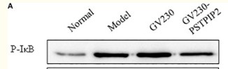
- Yao, Yao, et al. "PSTPIP2 inhibits the inflammatory response and proliferation of fibroblast-like synoviocytes in vitro." Frontiers in pharmacology 9 (2018): 1432.
-if-59.jpg)
- Immunofluorescence analysis of rat-spleen tissue. 1,IκB-α (phospho Ser32/S36) Polyclonal Antibody(red) was diluted at 1:200(4°C,overnight). 2, Cy3 labled Secondary antibody was diluted at 1:300(room temperature, 50min).3, Picture B: DAPI(blue) 10min. Picture A:Target. Picture B: DAPI. Picture C: merge of A+B
poly-ihc-rat-kidney.jpg)
- Immunohistochemical analysis of paraffin-embedded Rat-kidney tissue. 1,IκB-α (phospho Ser32/S36) Polyclonal Antibody was diluted at 1:200(4°C,overnight). 2, Sodium citrate pH 6.0 was used for antibody retrieval(>98°C,20min). 3,Secondary antibody was diluted at 1:200(room tempeRature, 30min). Negative control was used by secondary antibody only.
poly-ihc-mouse-kidney.jpg)
- Immunohistochemical analysis of paraffin-embedded Mouse-kidney tissue. 1,IκB-α (phospho Ser32/S36) Polyclonal Antibody was diluted at 1:200(4°C,overnight). 2, Sodium citrate pH 6.0 was used for antibody retrieval(>98°C,20min). 3,Secondary antibody was diluted at 1:200(room tempeRature, 30min). Negative control was used by secondary antibody only.
.jpg)
- Western Blot analysis of COS7 cells using Phospho-IκB-α (S32/S36) Polyclonal Antibody
- The picture was kindly provided by our customer.
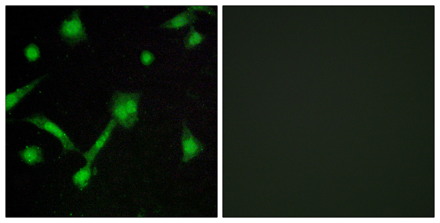
- Immunofluorescence analysis of LOVO cells, using IkappaB-alpha (Phospho-Ser32/Ser36) Antibody. The picture on the right is blocked with the phospho peptide.
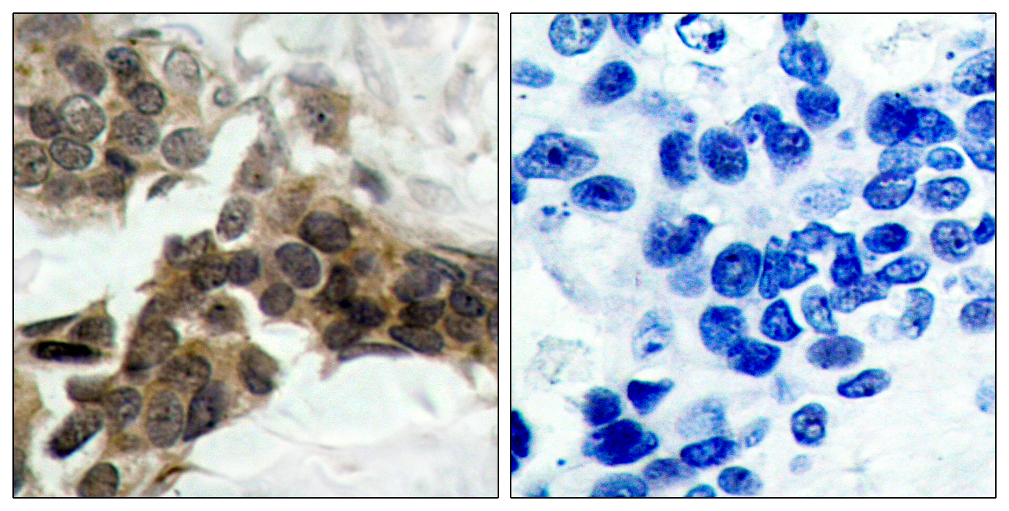
- Immunohistochemistry analysis of paraffin-embedded human breast carcinoma, using IkappaB-alpha (Phospho-Ser32/Ser36) Antibody. The picture on the right is blocked with the phospho peptide.
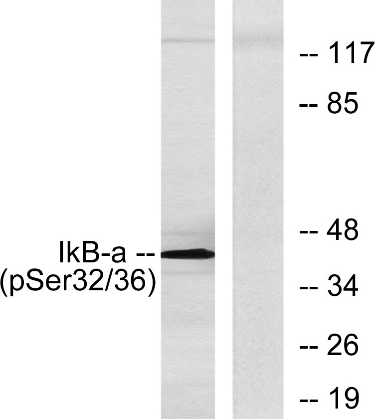
- Western blot analysis of lysates from COS7 cells, using IkappaB-alpha (Phospho-Ser32/Ser36) Antibody. The lane on the right is blocked with the phospho peptide.


