CD8 a (ABT304) Mouse mAb (Ready to Use)
- Catalog No.:YM6938R
- Applications:IHC
- Reactivity:Human
- Target:
- CD8
- Fields:
- >>Cell adhesion molecules;>>Antigen processing and presentation;>>Hematopoietic cell lineage;>>T cell receptor signaling pathway;>>Yersinia infection;>>Primary immunodeficiency
- Gene Name:
- CD8A MAL
- Protein Name:
- alpha polypeptide (p32);CD_antigen=CD8a;CD8;CD8 antigen alpha polypeptide;CD8 antigen alpha polypeptide (p32);CD8 antigen, alpha polypeptide (p32);CD8a;CD8A antigen;CD8A molecule;CD8A_HUMAN;Leu2;Leu2
- Human Swiss Prot No:
- P01732
- Mouse Swiss Prot No:
- P01731
- Rat Swiss Prot No:
- P07725
- Immunogen:
- Synthesized peptide derived from human CD8 AA range: 100-235
- Specificity:
- The antibody can specifically recognize human CD8 protein, including two typies of dimer: αβ heterodimer or αα homodimer.
- Formulation:
- PBS, pH7.2, 0.03% Porcolin 300, containing stabilizing protein
- Source:
- Monoclonal Mouse IgG2b, Kappa
- Dilution:
- Ready to use for IHC
- Purification:
- The antibody was affinity-purified from mouse ascites by affinity-chromatography using specific immunogen.
- Storage Stability:
- 2°C to 8°C/1 year
- Other Name:
- alpha polypeptide (p32);CD_antigen=CD8a;CD8;CD8 antigen alpha polypeptide;CD8 antigen alpha polypeptide (p32);CD8 antigen, alpha polypeptide (p32);CD8a;CD8A antigen;CD8A molecule;CD8A_HUMAN;Leu2;Leu2 T lymphocyte antigen;Ly 2;Ly 35;Ly B;Ly2;Ly3;Ly35;LyB;Lyt 2.1 lymphocyte differentiation antigen (AA at 100);LYT3;MAL;OKT8 T cell antigen;OTTHUMP00000160760;OTTHUMP00000160764;OTTHUMP00000203528;OTTHUMP00000203721;p32;T cell antigen Leu2;T cell co receptor;T lymphocyte differentiation antigen T8/Leu 2;T-cell surface glycoprotein CD8 alpha chain;T-cell surface glycoprotein Lyt 2;T-lymphocyte differentiation antigen T8/Leu-2;T8 T cell antigen;T8/Leu-2 T-lymphocyte differentiation antigen
- Molecular Weight(Da):
- 26kD
- Background:
- The CD8 antigen is a cell surface glycoprotein found on most cytotoxic T lymphocytes that mediates efficient cell-cell interactions within the immune system. The CD8 antigen acts as a coreceptor with the T-cell receptor on the T lymphocyte to recognize antigens displayed by an antigen presenting cell in the context of class I MHC molecules. The coreceptor functions as either a homodimer composed of two alpha chains or as a heterodimer composed of one alpha and one beta chain. Both alpha and beta chains share significant homology to immunoglobulin variable light chains. This gene encodes the CD8 alpha chain. Multiple transcript variants encoding different isoforms have been found for this gene. [provided by RefSeq, Nov 2011],
- Function:
- disease:Defects in CD8A are a cause of familial CD8 deficiency (CD8 deficiency) [MIM:608957]. Familial CD8 deficiency is a novel autosomal recessive immunologic defect characterized by absence of CD8+ cells, leading to recurrent bacterial infections.,function:Identifies cytotoxic/suppressor T-cells that interact with MHC class I bearing targets. CD8 is thought to play a role in the process of T-cell mediated killing. CD8 alpha chains binds to class I MHC molecules alpha-3 domains.,online information:CD8 entry,online information:CD8A mutation db,PTM:All of the five most carboxyl-terminal cysteines form inter-chain disulfide bonds in dimers and higher multimers, while the four N-terminal cysteines do not.,similarity:Contains 1 Ig-like V-type (immunoglobulin-like) domain.,subunit:In general heterodimer of an alpha and a beta chain linked by two disulfide bonds. Can also form homodimers. Sho
- Subcellular Location:
- Membranous
- Expression:
- Tonsil/ Appendix
- June 19-2018
- WESTERN IMMUNOBLOTTING PROTOCOL
- June 19-2018
- IMMUNOHISTOCHEMISTRY-PARAFFIN PROTOCOL
- June 19-2018
- IMMUNOFLUORESCENCE PROTOCOL
- September 08-2020
- FLOW-CYTOMEYRT-PROTOCOL
- May 20-2022
- Cell-Based ELISA│解您多样本WB检测之困扰
- July 13-2018
- CELL-BASED-ELISA-PROTOCOL-FOR-ACETYL-PROTEIN
- July 13-2018
- CELL-BASED-ELISA-PROTOCOL-FOR-PHOSPHO-PROTEIN
- July 13-2018
- Antibody-FAQs
- Products Images
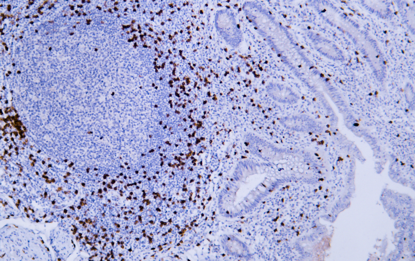
- Human appendix tissue was stained with Anti-CD8 (ABT304) Antibody
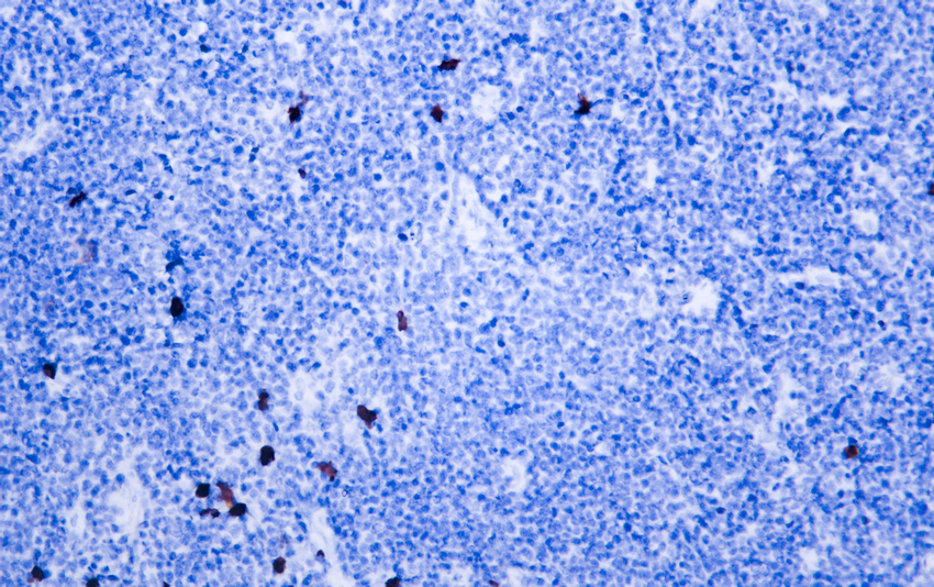
- Human burkitt lymphoma tissue was stained with Anti-CD8 (ABT304) Antibody
.jpg)
- Human lymphoma tissue was stained with Anti-CD8 (ABT304) Antibody
.jpg)
- Human lymphoma tissue was stained with Anti-CD8 (ABT304) Antibody

- Human tonsil tissue was stained with Anti-CD8 (ABT304) Antibody

- Fluorescence multiplex immunohistochemical analysis of Human tonsil tissue (formalin-fixed paraffin-embedded section). Merged staining of Anti-Podoplanin (YM6994), Anti-CD8 (YM6938), Anti-MCM2 (YM6077). The immunostaining was performed on a Leica Biosystems BOND® MAX instrument with an Sextuple-Fluorescence kit (RS0039, Immunoway). The section was incubated in 3 rounds of staining; sequentially for Anti-Podoplanin (YM6994 1:200), Anti-CD8 (YM6938 1:200), Anti-MCM2 (YM6077 1:200).; each using a separate fluorescent tyramide signal amplification system. EDTA based antigen retrieval (Leica Biosystems BOND® Epitope Retrieval Solution 2, pH 9.0, 20 minutes) was used in between rounds of tyramide signal amplification to remove the antibody from the previous round, to avoid any cross-reactivity. DAPI (dark blue) was used as a nuclear counter stain. Microscopy and pseudocoloring of individual dyes was performed using a Slideviewer Imaging System (3D histech).

- Fluorescence multiplex immunohistochemical analysis of Human tonsil tissue (formalin-fixed paraffin-embedded section). Merged staining of Anti-Podoplanin (YM6994), Anti-CD8 (YM6938), Anti-Ki-67 (YM6812). The immunostaining was performed on a Leica Biosystems BOND® MAX instrument with an Sextuple-Fluorescence kit (RS0039, Immunoway). The section was incubated in 3 rounds of staining; sequentially for Anti-Podoplanin (YM6994 1:200), Anti-CD8 (YM6938 1:200), Anti-Ki67 (YM6812 1:200).; each using a separate fluorescent tyramide signal amplification system. EDTA based antigen retrieval (Leica Biosystems BOND® Epitope Retrieval Solution 2, pH 9.0, 20 minutes) was used in between rounds of tyramide signal amplification to remove the antibody from the previous round, to avoid any cross-reactivity. DAPI (dark blue) was used as a nuclear counter stain. Microscopy and pseudocoloring of individual dyes was performed using a Slideviewer Imaging System (3D histech).

- Fluorescence multiplex immunohistochemical analysis of Human tonsil tissue (formalin-fixed paraffin-embedded section). Merged staining of Anti-Podoplanin (YM6994), Anti-CD8 (YM6938), Anti-MCM2 (YM6077). The immunostaining was performed on a Leica Biosystems BOND® MAX instrument with an Sextuple-Fluorescence kit (RS0039, Immunoway). The section was incubated in 3 rounds of staining; sequentially for Anti-Podoplanin (YM6994 1:200), Anti-CD8 (YM6938 1:200), Anti-MCM2 (YM6077 1:200).; each using a separate fluorescent tyramide signal amplification system. EDTA based antigen retrieval (Leica Biosystems BOND® Epitope Retrieval Solution 2, pH 9.0, 20 minutes) was used in between rounds of tyramide signal amplification to remove the antibody from the previous round, to avoid any cross-reactivity. DAPI (dark blue) was used as a nuclear counter stain. Microscopy and pseudocoloring of individual dyes was performed using a Slideviewer Imaging System (3D histech).


