α skeletal muscle actin Monoclonal Antibody(4B11)
- Catalog No.:YM3149
- Applications:WB;IHC;IF;IP
- Reactivity:Human;Mouse;Rat
- Target:
- Actin skeletal muscle α
- Gene Name:
- ACTA1
- Protein Name:
- Actin alpha skeletal muscle
- Human Gene Id:
- 58
- Human Swiss Prot No:
- P68133
- Mouse Gene Id:
- 11459
- Mouse Swiss Prot No:
- P68134
- Rat Gene Id:
- 29437
- Rat Swiss Prot No:
- P68136
- Immunogen:
- Synthetic Peptide of α skeletal muscle actin
- Specificity:
- The antibody detects endogenous α Skeletal Muscle Actin protein.
- Formulation:
- PBS, pH 7.4, containing 0.5%BSA, 0.02% sodium azide as Preservative and 50% Glycerol.
- Source:
- Monoclonal, Mouse
- Dilution:
- WB 1:500-10000 IP:1:200 IF 1:200 IHC 1:50-300
- Purification:
- The antibody was affinity-purified from mouse ascites by affinity-chromatography using specific immunogen.
- Storage Stability:
- -15°C to -25°C/1 year(Do not lower than -25°C)
- Other Name:
- ACTA1;ACTA;Actin, alpha skeletal muscle;Alpha-actin-1
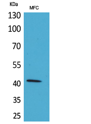
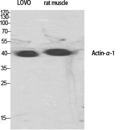
- Observed Band(KD):
- 42kD
- Background:
- The product encoded by this gene belongs to the actin family of proteins, which are highly conserved proteins that play a role in cell motility, structure and integrity. Alpha, beta and gamma actin isoforms have been identified, with alpha actins being a major constituent of the contractile apparatus, while beta and gamma actins are involved in the regulation of cell motility. This actin is an alpha actin that is found in skeletal muscle. Mutations in this gene cause nemaline myopathy type 3, congenital myopathy with excess of thin myofilaments, congenital myopathy with cores, and congenital myopathy with fiber-type disproportion, diseases that lead to muscle fiber defects. [provided by RefSeq, Jul 2008],
- Function:
- disease:Defects in ACTA1 are a cause of congenital myopathy with excess of thin myofilaments (CM) [MIM:102610].,disease:Defects in ACTA1 are a cause of congenital myopathy with fiber-type disproportion (CFTD) [MIM:255310]; also known as congenital fiber-type disproportion myopathy (CFTDM). CFTD is a genetically heterogeneous disorder in which there is relative hypotrophy of type 1 muscle fibers compared to type 2 fibers on skeletal muscle biopsy. However, these findings are not specific and can be found in many different myopathic and neuropathic conditions.,disease:Defects in ACTA1 are the cause of nemaline myopathy type 3 (NEM3) [MIM:161800]. Nemaline myopathy (NEM) is a form of congenital myopathy characterized by abnormal thread- or rod-like structures in muscle fibers on histologic examination. The clinical phenotype is highly variable, with differing age at onset and severity.,func
- Subcellular Location:
- Cytoplasm, cytoskeleton.
- Expression:
- Epithelium,Skeletal muscle,
- June 19-2018
- WESTERN IMMUNOBLOTTING PROTOCOL
- June 19-2018
- IMMUNOHISTOCHEMISTRY-PARAFFIN PROTOCOL
- June 19-2018
- IMMUNOFLUORESCENCE PROTOCOL
- September 08-2020
- FLOW-CYTOMEYRT-PROTOCOL
- May 20-2022
- Cell-Based ELISA│解您多样本WB检测之困扰
- July 13-2018
- CELL-BASED-ELISA-PROTOCOL-FOR-ACETYL-PROTEIN
- July 13-2018
- CELL-BASED-ELISA-PROTOCOL-FOR-PHOSPHO-PROTEIN
- July 13-2018
- Antibody-FAQs
- Products Images

- Immunohistochemical analysis of paraffin-embedded Rat-liver tissue. 1,α skeletal muscle actin Monoclonal Antibody(4B11) was diluted at 1:200(4°C,overnight). 2, Sodium citrate pH 6.0 was used for antibody retrieval(>98°C,20min). 3,Secondary antibody was diluted at 1:200(room tempeRature, 30min). Negative control was used by secondary antibody only.
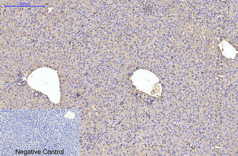
- Immunohistochemical analysis of paraffin-embedded Mouse-liver tissue. 1,α skeletal muscle actin Monoclonal Antibody(4B11) was diluted at 1:200(4°C,overnight). 2, Sodium citrate pH 6.0 was used for antibody retrieval(>98°C,20min). 3,Secondary antibody was diluted at 1:200(room tempeRature, 30min). Negative control was used by secondary antibody only.
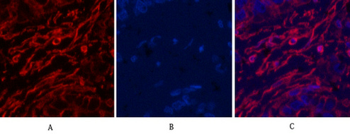
- Immunofluorescence analysis of Human-liver-cancer tissue. 1,α skeletal muscle actin Monoclonal Antibody(4B11)(red) was diluted at 1:200(4°C,overnight). 2, Cy3 labled Secondary antibody was diluted at 1:300(room temperature, 50min).3, Picture B: DAPI(blue) 10min. Picture A:Target. Picture B: DAPI. Picture C: merge of A+B
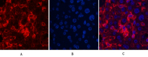
- Immunofluorescence analysis of Mouse-liver tissue. 1,α skeletal muscle actin Monoclonal Antibody(4B11)(red) was diluted at 1:200(4°C,overnight). 2, Cy3 labled Secondary antibody was diluted at 1:300(room temperature, 50min).3, Picture B: DAPI(blue) 10min. Picture A:Target. Picture B: DAPI. Picture C: merge of A+B
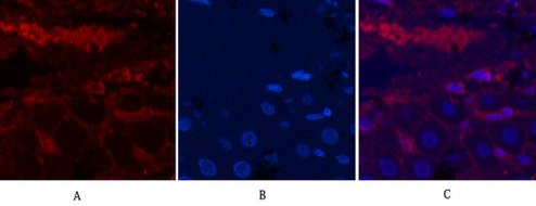
- Immunofluorescence analysis of Rat-liver tissue. 1,α skeletal muscle actin Monoclonal Antibody(4B11)(red) was diluted at 1:200(4°C,overnight). 2, Cy3 labled Secondary antibody was diluted at 1:300(room temperature, 50min).3, Picture B: DAPI(blue) 10min. Picture A:Target. Picture B: DAPI. Picture C: merge of A+B
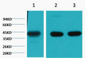
- Western blot analysis of 1) Hela, 2) Mouse Brain tissue, 3) Rat Brain tissue, diluted at 1:20000.
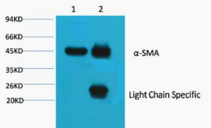
- 1) Input: Mouse Brain Tissue Lysate 2) IP product: IP dilute 1: 200
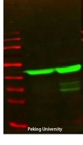
- The picture was kindly provided by our customer