CD1 Monoclonal Antibody(9H6)
- Catalog No.:YM3068
- Applications:IHC;IF
- Reactivity:Human;Mouse;Rat
- Target:
- CD1
- Fields:
- >>Tight junction;>>Hematopoietic cell lineage;>>Amoebiasis
- Gene Name:
- CD1A
- Protein Name:
- T-cell surface glycoprotein CD1a
- Human Gene Id:
- 909
- Human Swiss Prot No:
- P06126
- Immunogen:
- Synthetic Peptide of CD1
- Specificity:
- The antibody detects endogenous CD1 proteins.
- Formulation:
- PBS, pH 7.4, containing 0.5%BSA, 0.02% sodium azide as Preservative and 50% Glycerol.
- Source:
- Monoclonal, Mouse
- Dilution:
- IHC 1:50-200. IF 1:50-200
- Purification:
- The antibody was affinity-purified from mouse ascites by affinity-chromatography using specific immunogen.
- Storage Stability:
- -15°C to -25°C/1 year(Do not lower than -25°C)
- Other Name:
- T-cell surface glycoprotein CD1a (T-cell surface antigen T6/Leu-6) (hTa1 thymocyte antigen) (CD antigen CD1a)
- Background:
- This gene encodes a member of the CD1 family of transmembrane glycoproteins, which are structurally related to the major histocompatibility complex (MHC) proteins and form heterodimers with beta-2-microglobulin. The CD1 proteins mediate the presentation of primarily lipid and glycolipid antigens of self or microbial origin to T cells. The human genome contains five CD1 family genes organized in a cluster on chromosome 1. The CD1 family members are thought to differ in their cellular localization and specificity for particular lipid ligands. The protein encoded by this gene localizes to the plasma membrane and to recycling vesicles of the early endocytic system. Alternative splicing results in multiple transcript variants. [provided by RefSeq, Mar 2016],
- Function:
- function:Antigen-presenting protein that binds self and non-self lipid and glycolipid antigens and presents them to T-cell receptors on natural killer T-cells.,miscellaneous:During protein synthesis and maturation, CD1 family members bind endogenous lipids that are replaced by lipid or glycolipid antigens when the proteins are internalized and pass through endosomes, before trafficking back to the cell surface.,similarity:Contains 1 Ig-like (immunoglobulin-like) domain.,subcellular location:Subject to intracellular trafficking between the cell membrane and endosomes. Localizes to cell surface lipid rafts.,subunit:Heterodimer with B2M (beta-2-microglobulin). Interacts with CD74.,tissue specificity:Expressed on cortical thymocytes, epidermal Langerhans cells, dendritic cells, on certain T-cell leukemias, and in various other tissues.,
- Subcellular Location:
- Cell membrane ; Single-pass type I membrane protein . Membrane raft ; Single-pass type I membrane protein . Endosome membrane ; Single-pass type I membrane protein . Subject to intracellular trafficking between the cell membrane and endosomes (PubMed:11231314). Localizes to cell surface lipid rafts (PubMed:18178838). .
- Expression:
- Expressed on cortical thymocytes, epidermal Langerhans cells, dendritic cells, on certain T-cell leukemias, and in various other tissues.
- June 19-2018
- WESTERN IMMUNOBLOTTING PROTOCOL
- June 19-2018
- IMMUNOHISTOCHEMISTRY-PARAFFIN PROTOCOL
- June 19-2018
- IMMUNOFLUORESCENCE PROTOCOL
- September 08-2020
- FLOW-CYTOMEYRT-PROTOCOL
- May 20-2022
- Cell-Based ELISA│解您多样本WB检测之困扰
- July 13-2018
- CELL-BASED-ELISA-PROTOCOL-FOR-ACETYL-PROTEIN
- July 13-2018
- CELL-BASED-ELISA-PROTOCOL-FOR-PHOSPHO-PROTEIN
- July 13-2018
- Antibody-FAQs
- Products Images
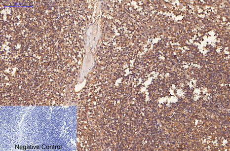
- Immunohistochemical analysis of paraffin-embedded Human-Tonsil tissue. 1,CD1 Monoclonal Antibody(9H6) was diluted at 1:200(4°C,overnight). 2, Sodium citrate pH 6.0 was used for antibody retrieval(>98°C,20min). 3,Secondary antibody was diluted at 1:200(room tempeRature, 30min). Negative control was used by secondary antibody only.
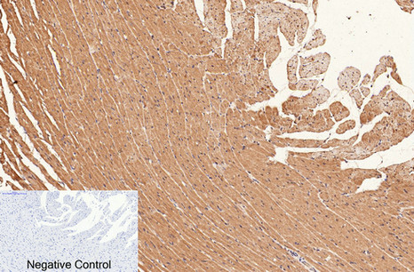
- Immunohistochemical analysis of paraffin-embedded Rat-heart tissue. 1,CD1 Monoclonal Antibody(9H6) was diluted at 1:200(4°C,overnight). 2, Sodium citrate pH 6.0 was used for antibody retrieval(>98°C,20min). 3,Secondary antibody was diluted at 1:200(room tempeRature, 30min). Negative control was used by secondary antibody only.

- Immunohistochemical analysis of paraffin-embedded Mouse-heart tissue. 1,CD1 Monoclonal Antibody(9H6) was diluted at 1:200(4°C,overnight). 2, Sodium citrate pH 6.0 was used for antibody retrieval(>98°C,20min). 3,Secondary antibody was diluted at 1:200(room tempeRature, 30min). Negative control was used by secondary antibody only.

- Immunofluorescence analysis of Mouse-heart tissue. 1,CD1 Monoclonal Antibody(9H6)(red) was diluted at 1:200(4°C,overnight). 2, Cy3 labled Secondary antibody was diluted at 1:300(room temperature, 50min).3, Picture B: DAPI(blue) 10min. Picture A:Target. Picture B: DAPI. Picture C: merge of A+B


