HER2 Monoclonal Antibody(11H9)
- Catalog No.:YM3045
- Applications:WB;IF;IHC
- Reactivity:Human;Mouse;Rat
- Target:
- HER2
- Fields:
- >>EGFR tyrosine kinase inhibitor resistance;>>Endocrine resistance;>>Platinum drug resistance;>>MAPK signaling pathway;>>ErbB signaling pathway;>>Calcium signaling pathway;>>HIF-1 signaling pathway;>>PI3K-Akt signaling pathway;>>Focal adhesion;>>Adherens junction;>>Tight junction;>>Pathways in cancer;>>Proteoglycans in cancer;>>MicroRNAs in cancer;>>Pancreatic cancer;>>Endometrial cancer;>>Prostate cancer;>>Bladder cancer;>>Non-small cell lung cancer;>>Breast cancer;>>Gastric cancer;>>Central carbon metabolism in cancer
- Gene Name:
- ERBB2
- Protein Name:
- Receptor tyrosine-protein kinase erbB-2
- Human Gene Id:
- 2064
- Human Swiss Prot No:
- P04626
- Mouse Gene Id:
- 13866
- Mouse Swiss Prot No:
- P70424
- Rat Swiss Prot No:
- P06494
- Immunogen:
- Synthetic Peptide of HER2
- Specificity:
- The antibody detects endogenous ErbB-2/HER-2 proteins.
- Formulation:
- PBS, pH 7.4, containing 0.5%BSA, 0.02% sodium azide as Preservative and 50% Glycerol.
- Source:
- Monoclonal, Mouse
- Dilution:
- WB 1:2000-4000 IHC 1:200 IF 1:200
- Purification:
- The antibody was affinity-purified from mouse ascites by affinity-chromatography using specific immunogen.
- Storage Stability:
- -15°C to -25°C/1 year(Do not lower than -25°C)
- Other Name:
- ERBB2;HER2;MLN19;NEU;NGL;Receptor tyrosine-protein kinase erbB-2;Metastatic lymph node gene 19 protein;MLN 19;Proto-oncogene Neu;Proto-oncogene c-ErbB-2;Tyrosine kinase-type cell surface receptor HER2;p185erbB2;CD340
- Observed Band(KD):
- 180kD
- Background:
- This gene encodes a member of the epidermal growth factor (EGF) receptor family of receptor tyrosine kinases. This protein has no ligand binding domain of its own and therefore cannot bind growth factors. However, it does bind tightly to other ligand-bound EGF receptor family members to form a heterodimer, stabilizing ligand binding and enhancing kinase-mediated activation of downstream signalling pathways, such as those involving mitogen-activated protein kinase and phosphatidylinositol-3 kinase. Allelic variations at amino acid positions 654 and 655 of isoform a (positions 624 and 625 of isoform b) have been reported, with the most common allele, Ile654/Ile655, shown here. Amplification and/or overexpression of this gene has been reported in numerous cancers, including breast and ovarian tumors. Alternative splicing results in several additional transcript variants, some encoding d
- Function:
- catalytic activity:ATP + a [protein]-L-tyrosine = ADP + a [protein]-L-tyrosine phosphate.,disease:Defects in ERBB2 are associated with familial glioma of brain [MIM:137800]; also called glioblastoma multiforme. Gliomas are central nervous system neoplasms derived from glial cells and comprise astrocytomas, glioblastoma multiforme, oligodendrogliomas, and ependymomas.,disease:Defects in ERBB2 are associated with gastric cancer [MIM:137215]; also known as hereditary familial diffuse gastric cancer (HDGC).,disease:Defects in ERBB2 are associated with lung cancer [MIM:211980]; also called adenocarcinoma of lung.,disease:Defects in ERBB2 are associated with ovarian cancer [MIM:167000]. Ovarian cancer is the leading cause of death from gynecologic malignancy. It is characterized by advanced presentation with loco-regional dissemination in the peritoneal cavity and the rare incidence of viscera
- Subcellular Location:
- [Isoform 1]: Cell membrane ; Single-pass type I membrane protein. Early endosome . Cytoplasm, perinuclear region. Nucleus. Translocation to the nucleus requires endocytosis, probably endosomal sorting and is mediated by importin beta-1/KPNB1. Also detected in VPS35-positive endosome-to-TGN retrograde vesicles (PubMed:31138794). .; [Isoform 2]: Cytoplasm. Nucleus.; [Isoform 3]: Cytoplasm. Nucleus.
- Expression:
- Expressed in a variety of tumor tissues including primary breast tumors and tumors from small bowel, esophagus, kidney and mouth.
HSP90 inhibitor AUY922 can reverse Fulvestrant induced feedback reaction in human breast cancer cells. CANCER SCIENCE 2017 May 19 WB Human MCF-7 cell,T47D cell
BMP9 Inhibits Proliferation and Metastasis of HER2-Positive SK-BR-3 Breast Cancer Cells through ERK1/2 and PI3K/AKT Pathways. PLoS One 2014 May 07 WB,IHC Mouse,Human 1:500 SK-BR-3 cell-xenograft SK-BR-3 cell,MDA-MB-231 cell
Pyrotinib treatment on HER2-positive gastric cancer cells promotes the released exosomes to enhance endothelial cell progression, which can be counteracted by apatinib. OncoTargets and Therapy Oncotargets Ther. 2019; 12: 2777–2787 WB Human 1:1000 SGC7901 cell, MGC-803 cell, BGC823 cell, AGS cell,MKN45 cell,NCI-N87 cell
A novel multi-target inhibitor harboring selectivity of inhibiting EGFR T790M sparing wild-type EGFR. American Journal of Cancer Research Am J Cancer Res. 2017; 7(9): 1884–1898 WB Human NCI-H1975 cell,A549 cell
Contribution of PGAP3 co-amplified and co-overexpressed with ERBB2 at 17q12 involved poor prognosis in gastric cancer. Jingcui Yu WB Human 1:1000 NCI-N87 cell
GDF-15 Inhibits ADP-Induced Human Platelet Aggregation through the GFRAL/RET Signaling Complex Biomolecules Baikang Xie WB Human 1:1000 platelets,erythrocytes,leukocytes
- June 19-2018
- WESTERN IMMUNOBLOTTING PROTOCOL
- June 19-2018
- IMMUNOHISTOCHEMISTRY-PARAFFIN PROTOCOL
- June 19-2018
- IMMUNOFLUORESCENCE PROTOCOL
- September 08-2020
- FLOW-CYTOMEYRT-PROTOCOL
- May 20-2022
- Cell-Based ELISA│解您多样本WB检测之困扰
- July 13-2018
- CELL-BASED-ELISA-PROTOCOL-FOR-ACETYL-PROTEIN
- July 13-2018
- CELL-BASED-ELISA-PROTOCOL-FOR-PHOSPHO-PROTEIN
- July 13-2018
- Antibody-FAQs
- Products Images
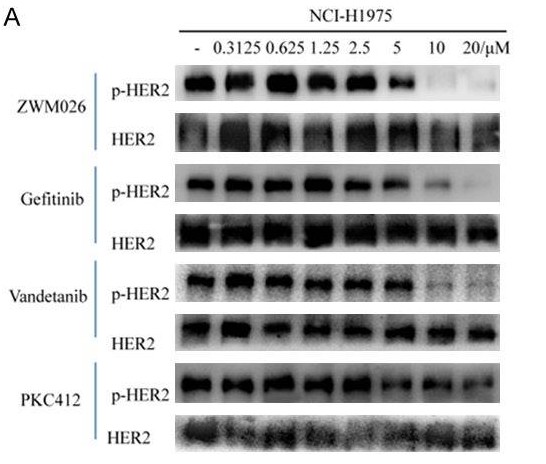
- Song, Xiaoping, et al. "A novel multi-target inhibitor harboring selectivity of inhibiting egfr T790M sparing wild-type EGFR." American journal of cancer research 7.9 (2017): 1884.
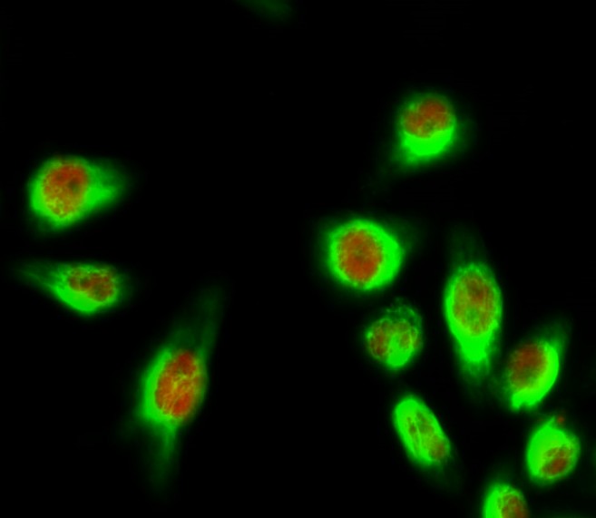
- Immunofluorescence analysis of Hela cell. 1,E2F-1 Polyclonal Antibody(red) was diluted at 1:200(4° overnight). HER2 Monoclonal Antibody(11H9)(green) was diluted at 1:200(4° overnight). 2, Goat Anti Rabbit Alexa Fluor 594 Catalog:RS3611 was diluted at 1:1000(room temperature, 50min). Goat Anti Mouse Alexa Fluor 488 Catalog:RS3208 was diluted at 1:1000(room temperature, 50min).
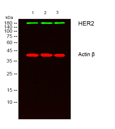
- Western blot analysis of lysates from 1) Hela, 2) A431,3) MCF-7 cells, (Green) primary antibody was diluted at 1:1000, 4°over night, secondary antibody(cat:RS23910)was diluted at 1:10000, 37° 1hour. (Red) Actin β Polyclonal Antibody (cat:YT0099) antibody was diluted at 1:5000 as loading control, 4° over night,secondary antibody(cat:RS23720)was diluted at 1:10000, 37° 1hour.

- Immunohistochemical analysis of paraffin-embedded Human-Tonsil tissue. 1,HER2 Monoclonal Antibody(11H9) was diluted at 1:200(4°C,overnight). 2, Sodium citrate pH 6.0 was used for antibody retrieval(>98°C,20min). 3,Secondary antibody was diluted at 1:200(room tempeRature, 30min). Negative control was used by secondary antibody only.
- Immunohistochemical analysis of paraffin-embedded Rat-kidney tissue. 1,HER2 Monoclonal Antibody(11H9) was diluted at 1:200(4°C,overnight). 2, Sodium citrate pH 6.0 was used for antibody retrieval(>98°C,20min). 3,Secondary antibody was diluted at 1:200(room tempeRature, 30min). Negative control was used by secondary antibody only.

- Immunohistochemical analysis of paraffin-embedded Mouse-testis tissue. 1,HER2 Monoclonal Antibody(11H9) was diluted at 1:200(4°C,overnight). 2, Sodium citrate pH 6.0 was used for antibody retrieval(>98°C,20min). 3,Secondary antibody was diluted at 1:200(room tempeRature, 30min). Negative control was used by secondary antibody only.
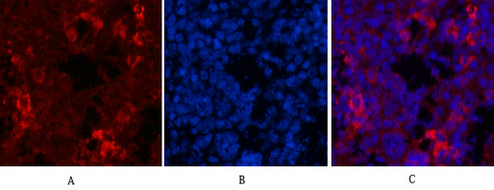
- Immunofluorescence analysis of Mouse-spleen tissue. 1,HER2 Monoclonal Antibody(11H9)(red) was diluted at 1:200(4°C,overnight). 2, Cy3 labled Secondary antibody was diluted at 1:300(room temperature, 50min).3, Picture B: DAPI(blue) 10min. Picture A:Target. Picture B: DAPI. Picture C: merge of A+B
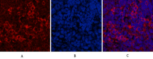
- Immunofluorescence analysis of Rat-spleen tissue. 1,HER2 Monoclonal Antibody(11H9)(red) was diluted at 1:200(4°C,overnight). 2, Cy3 labled Secondary antibody was diluted at 1:300(room temperature, 50min).3, Picture B: DAPI(blue) 10min. Picture A:Target. Picture B: DAPI. Picture C: merge of A+B

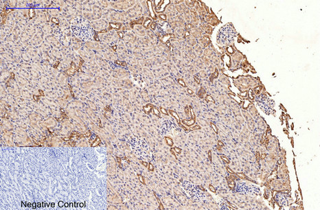
- IHC staining of human breast cancer tissue, diluted at 1:200.

- Western blot analysis of lysates from 1) Hela, 2) A431,3) MCF-7 cells, (Green) primary antibody was diluted at 1:1000, 4°over night, secondary antibody(cat:RS23910)was diluted at 1:10000, 37° 1hour. (Red) Actin β Polyclonal Antibody (cat:YT0099) antibody was diluted at 1:5000 as loading control, 4° over night,secondary antibody(cat:RS23720)was diluted at 1:10000, 37° 1hour.


