HP1-γ mouse mAb
- Catalog No.:YM1216
- Applications:WB;IF;IP;IHC
- Reactivity:Mouse;Human;Monkey;Hamster;Rat
- Target:
- HP1γ
- Fields:
- >>Shigellosis
- Gene Name:
- cbx3
- Human Gene Id:
- 11335
- Human Swiss Prot No:
- Q13185
- Mouse Swiss Prot No:
- P23198
- Immunogen:
- Purified recombinant human HP1-gamma protein fragments expressed in E.coli.
- Specificity:
- This antibody detects endogenous levels of HP1-gamma and does not cross-react with related proteins.
- Formulation:
- Liquid in PBS containing 50% glycerol, 0.5% BSA and 0.02% sodium azide.
- Source:
- Monoclonal, Mouse
- Dilution:
- wb 1:1000 icc 1:200. IF 1:50-200
- Purification:
- The antibody was affinity-purified from mouse ascites by affinity-chromatography using epitope-specific immunogen.
- Concentration:
- 1 mg/ml
- Storage Stability:
- -15°C to -25°C/1 year(Do not lower than -25°C)
- Other Name:
- CBX 3;CBX3;CBX3_HUMAN;Chromobox homolog 3 (HP1 gamma homolog, Drosophila);Chromobox homolog 3;Chromobox protein homolog 3;GAMMA;HECH;Heterochromatin like protein 1;Heterochromatin protein 1 homolog gamma;Heterochromatin protein HP1 gamma;HP1 gamma;HP1 gamma homolog;HP1Hs gamma;Modifier 2 protein.
- Observed Band(KD):
- 22kD
- Background:
- At the nuclear envelope, the nuclear lamina and heterochromatin are adjacent to the inner nuclear membrane. The protein encoded by this gene binds DNA and is a component of heterochromatin. This protein also can bind lamin B receptor, an integral membrane protein found in the inner nuclear membrane. The dual binding functions of the encoded protein may explain the association of heterochromatin with the inner nuclear membrane. This protein binds histone H3 tails methylated at Lys-9 sites. This protein is also recruited to sites of ultraviolet-induced DNA damage and double-strand breaks. Two transcript variants encoding the same protein but differing in the 5' UTR, have been found for this gene.[provided by RefSeq, Mar 2011],
- Function:
- function:Seems to be involved in transcriptional silencing in heterochromatin-like complexes. Recognizes and binds histone H3 tails methylated at 'Lys-9', leading to epigenetic repression. May contribute to the association of the heterochromatin with the inner nuclear membrane through its interaction with lamin B receptor (LBR). Involved in the formation of functional kinetochore through interaction with MIS12 complex proteins.,PTM:Phosphorylated by PIM1. Phosphorylated during interphase and possibly hyper-phosphorylated during mitosis.,similarity:Contains 2 chromo domains.,subcellular location:Associates with euchromatin and is largely excluded from constitutive heterochromatin. May be associated with microtubules and mitotic poles during mitosis.,subunit:Binds directly to CHAF1A. Interacts with histone H3 methylated at 'Lys-9'. Part of the E2F6.com-1 complex in G0 phase composed of E2F
- Subcellular Location:
- Nucleus . Associates with euchromatin and is largely excluded from constitutive heterochromatin. May be associated with microtubules and mitotic poles during mitosis (Potential). .
- Expression:
- Bone marrow,Brain,Cajal-Retzius cell,Epithelium,Liver,Placenta,
- June 19-2018
- WESTERN IMMUNOBLOTTING PROTOCOL
- June 19-2018
- IMMUNOHISTOCHEMISTRY-PARAFFIN PROTOCOL
- June 19-2018
- IMMUNOFLUORESCENCE PROTOCOL
- September 08-2020
- FLOW-CYTOMEYRT-PROTOCOL
- May 20-2022
- Cell-Based ELISA│解您多样本WB检测之困扰
- July 13-2018
- CELL-BASED-ELISA-PROTOCOL-FOR-ACETYL-PROTEIN
- July 13-2018
- CELL-BASED-ELISA-PROTOCOL-FOR-PHOSPHO-PROTEIN
- July 13-2018
- Antibody-FAQs
- Products Images
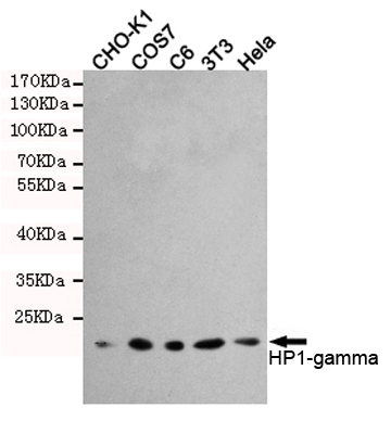
- Western blot detection of HP1-gamma in Hela,3T3,C6,COS7 and CHO-K1 cell lysates using HP1-gamma mouse mAb (1:1000 diluted).Predicted band size:22KDa.Observed band size:22KDa.
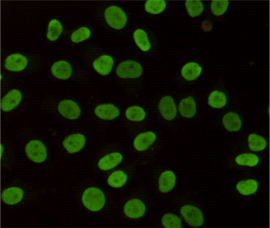
- Immunocytochemistry staining of HeLa cells fixed with 4% Paraformaldehyde and using anti-HP1-gamma mouse mAb (dilution 1:200).
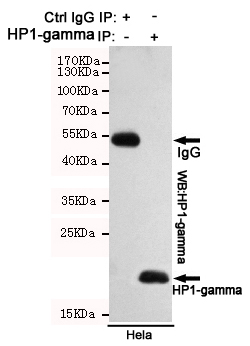
- Immunoprecipitation analysis of Hela cell lysates using HP1-gamma mouse mAb.
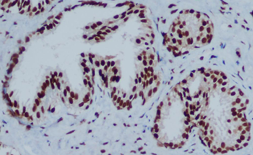
- Immunohistochemical analysis of paraffin-embedded Prostate Cancer using HP1-gamma mouse mAb (1/200 dilution).Antigen retrieval was performed by pressure cooking in citrate buffer (pH 6.0).
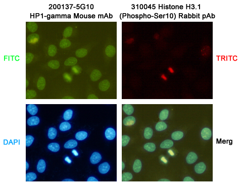
- Immunocytochemistry staining of HeLa cells fixed with -20℃ Methanol and using HP1-gamma (200137-5G10,dilution 1:200) mouse mAb (green) and Histone H3.1 (Phospho-Ser10)(310045,dilution 1:200) Rabbit pAb (red). DAPI was used to stain nucleus(blue).


