DCNL3 rabbit pAb
- Catalog No.:YT7767
- Applications:WB
- Reactivity:Human;Mouse;Rat
- Target:
- DCNL3
- Gene Name:
- DCUN1D3
- Protein Name:
- DCNL3
- Human Gene Id:
- 123879
- Human Swiss Prot No:
- Q8IWE4
- Mouse Gene Id:
- 233805
- Mouse Swiss Prot No:
- Q8K0V2
- Rat Gene Id:
- 309035
- Rat Swiss Prot No:
- Q4V8B2
- Immunogen:
- Synthesized peptide derived from human DCNL3 AA range: 205-255
- Specificity:
- This antibody detects endogenous levels of DCNL3 at Human/Mouse/Rat
- Formulation:
- Liquid in PBS containing 50% glycerol, 0.5% BSA and 0.02% sodium azide.
- Source:
- Polyclonal, Rabbit,IgG
- Dilution:
- WB 1:500-2000
- Purification:
- The antibody was affinity-purified from rabbit antiserum by affinity-chromatography using epitope-specific immunogen.
- Concentration:
- 1 mg/ml
- Storage Stability:
- -15°C to -25°C/1 year(Do not lower than -25°C)
- Molecular Weight(Da):
- 33kD
- Function:
- similarity:Contains 1 DCUN1 domain.,
- Subcellular Location:
- Cell membrane . Cytoplasm . Nucleus . Cytoplasm, perinuclear region . After UVC treatment, the protein enters to the nucleus gradually (PubMed:18823379). Cell membrane localization is essential for CUL3 neddylation (PubMed:19617556). .
- Expression:
- Tends to be down-regulated in different type of cancers, including lung neuroendocrine carcinoma, thyroid Huerthle cell carcinoma and lung squamous cell carcinoma (PubMed:25349211). Mostly expressed in testis and brain (PubMed:26906416). Highly expressed in liver, bladder and renal normal tissue than their tumor tissue counterparts (PubMed:18823379). Palmitoylation stabilizes DCUN1D3 at the cell membrane (PubMed:19617556).
- June 19-2018
- WESTERN IMMUNOBLOTTING PROTOCOL
- June 19-2018
- IMMUNOHISTOCHEMISTRY-PARAFFIN PROTOCOL
- June 19-2018
- IMMUNOFLUORESCENCE PROTOCOL
- September 08-2020
- FLOW-CYTOMEYRT-PROTOCOL
- May 20-2022
- Cell-Based ELISA│解您多样本WB检测之困扰
- July 13-2018
- CELL-BASED-ELISA-PROTOCOL-FOR-ACETYL-PROTEIN
- July 13-2018
- CELL-BASED-ELISA-PROTOCOL-FOR-PHOSPHO-PROTEIN
- July 13-2018
- Antibody-FAQs
- Products Images
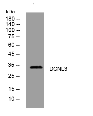
- Western blot analysis of lysates from HpeG2 cells, primary antibody was diluted at 1:1000, 4°over night


