NGFR p75 Polyclonal Antibody
- Catalog No.:YT3116
- Applications:WB;IF;ELISA
- Reactivity:Human;Mouse;Rat
- Target:
- NGFR p75
- Fields:
- >>MAPK signaling pathway;>>Ras signaling pathway;>>Rap1 signaling pathway;>>Cytokine-cytokine receptor interaction;>>PI3K-Akt signaling pathway;>>Apoptosis - multiple species;>>Neurotrophin signaling pathway;>>Transcriptional misregulation in cancer
- Gene Name:
- NGFR
- Protein Name:
- Tumor necrosis factor receptor superfamily member 16
- Human Gene Id:
- 4804
- Human Swiss Prot No:
- P08138
- Mouse Swiss Prot No:
- Q9Z0W1
- Rat Gene Id:
- 24596
- Rat Swiss Prot No:
- P07174
- Immunogen:
- The antiserum was produced against synthesized peptide derived from human TNR16. AA range:121-170
- Specificity:
- NGFR p75 Polyclonal Antibody detects endogenous levels of NGFR p75 protein.
- Formulation:
- Liquid in PBS containing 50% glycerol, 0.5% BSA and 0.02% sodium azide.
- Source:
- Polyclonal, Rabbit,IgG
- Dilution:
- WB 1:500 - 1:2000. IF 1:200 - 1:1000. ELISA: 1:40000. Not yet tested in other applications.
- Purification:
- The antibody was affinity-purified from rabbit antiserum by affinity-chromatography using epitope-specific immunogen.
- Concentration:
- 1 mg/ml
- Storage Stability:
- -15°C to -25°C/1 year(Do not lower than -25°C)
- Other Name:
- NGFR;TNFRSF16;Tumor necrosis factor receptor superfamily member 16;Gp80-LNGFR;Low affinity neurotrophin receptor p75NTR;Low-affinity nerve growth factor receptor;NGF receptor;p75 ICD;CD antigen CD271
- Observed Band(KD):
- 75kD
- Background:
- Nerve growth factor receptor contains an extracellular domain containing four 40-amino acid repeats with 6 cysteine residues at conserved positions followed by a serine/threonine-rich region, a single transmembrane domain, and a 155-amino acid cytoplasmic domain. The cysteine-rich region contains the nerve growth factor binding domain. [provided by RefSeq, Jul 2008],
- Function:
- domain:Death domain is responsible for interaction with RANBP9.,domain:The extracellular domain is responsible for interaction with NTRK1.,function:Low affinity receptor which can bind to NGF, BDNF, NT-3, and NT-4. Can mediate cell survival as well as cell death of neural cells.,PTM:N- and O-glycosylated.,PTM:O-linked glycans consist of Gal(1-3)GalNAc core elongated by 1 or 2 NeuNAc.,PTM:Phosphorylated on serine residues.,similarity:Contains 1 death domain.,similarity:Contains 4 TNFR-Cys repeats.,subunit:Homodimer; disulfide-linked. Interacts with p75NTR-associated cell death executor. Interacts with TRAF2, TRAF4, TRAF6, PTPN13 and RANBP9. Interacts through TRAF6 with SQSTM1 which bridges NGFR to NTRK1. Interacts with BEX1 and NGFRAP1/BEX3. Interacts with KIDINS220 and NTRK1. Can form a ternary complex with NTRK1 and KIDINS220 and this complex is affected by the expression levels of KIDI
- Subcellular Location:
- Cell membrane ; Single-pass type I membrane protein . Perikaryon . Cell projection, growth cone . Cell projection, dendritic spine .
- Expression:
- Brain,
IFT80 Improves Invasion Ability in Gastric Cancer Cell Line via ift80/p75NGFR/MMP9 Signaling. INTERNATIONAL JOURNAL OF MOLECULAR SCIENCES 2018 Nov 16 WB Human 1:1000 SGC-7901 cell
- June 19-2018
- WESTERN IMMUNOBLOTTING PROTOCOL
- June 19-2018
- IMMUNOHISTOCHEMISTRY-PARAFFIN PROTOCOL
- June 19-2018
- IMMUNOFLUORESCENCE PROTOCOL
- September 08-2020
- FLOW-CYTOMEYRT-PROTOCOL
- May 20-2022
- Cell-Based ELISA│解您多样本WB检测之困扰
- July 13-2018
- CELL-BASED-ELISA-PROTOCOL-FOR-ACETYL-PROTEIN
- July 13-2018
- CELL-BASED-ELISA-PROTOCOL-FOR-PHOSPHO-PROTEIN
- July 13-2018
- Antibody-FAQs
- Products Images
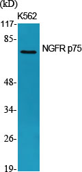
- Western Blot analysis of various cells using NGFR p75 Polyclonal Antibody
.jpg)
- Western Blot analysis of HepG2 cells using NGFR p75 Polyclonal Antibody

- Immunofluorescence analysis of A549 cells, using TNR16 Antibody. The picture on the right is blocked with the synthesized peptide.

- Western blot analysis of lysates from HepG2 cells, using TNR16 Antibody. The lane on the right is blocked with the synthesized peptide.

- Western blot analysis of the lysates from HeLa cells using TNR16 antibody.


