FoxO3A Polyclonal Antibody
- Catalog No.:YT1763
- Applications:WB;IHC;IF;ELISA
- Reactivity:Human;Mouse;Rat
- Target:
- FoxO3A
- Fields:
- >>EGFR tyrosine kinase inhibitor resistance;>>Chemokine signaling pathway;>>FoxO signaling pathway;>>Mitophagy - animal;>>PI3K-Akt signaling pathway;>>AMPK signaling pathway;>>Longevity regulating pathway;>>Longevity regulating pathway - multiple species;>>Cellular senescence;>>Neurotrophin signaling pathway;>>Prolactin signaling pathway;>>Alcoholic liver disease;>>Shigellosis;>>Chemical carcinogenesis - reactive oxygen species;>>Endometrial cancer;>>Non-small cell lung cancer
- Gene Name:
- FOXO3
- Protein Name:
- Forkhead box protein O3
- Human Gene Id:
- 2309
- Human Swiss Prot No:
- O43524
- Mouse Gene Id:
- 56484
- Mouse Swiss Prot No:
- Q9WVH4
- Immunogen:
- The antiserum was produced against synthesized peptide derived from human FKHRL1. AA range:220-269
- Specificity:
- FoxO3A Polyclonal Antibody detects endogenous levels of FoxO3A protein.
- Formulation:
- Liquid in PBS containing 50% glycerol, 0.5% BSA and 0.02% sodium azide.
- Source:
- Polyclonal, Rabbit,IgG
- Dilution:
- WB 1:500 - 1:2000. IHC 1:100 - 1:300. IF 1:200 - 1:1000. ELISA: 1:20000. Not yet tested in other applications.
- Purification:
- The antibody was affinity-purified from rabbit antiserum by affinity-chromatography using epitope-specific immunogen.
- Concentration:
- 1 mg/ml
- Storage Stability:
- -15°C to -25°C/1 year(Do not lower than -25°C)
- Other Name:
- FOXO3;FKHRL1;FOXO3A;Forkhead box protein O3;AF6q21 protein;Forkhead in rhabdomyosarcoma-like 1
- Observed Band(KD):
- 90kD
- Background:
- This gene belongs to the forkhead family of transcription factors which are characterized by a distinct forkhead domain. This gene likely functions as a trigger for apoptosis through expression of genes necessary for cell death. Translocation of this gene with the MLL gene is associated with secondary acute leukemia. Alternatively spliced transcript variants encoding the same protein have been observed. [provided by RefSeq, Jul 2008],
- Function:
- disease:A chromosomal aberration involving FOXO3 is found in secondary acute leukemias. Translocation t(6;11)(q21;q23) with MLL/HRX.,function:Transcriptional activator which triggers apoptosis in the absence of survival factors, including neuronal cell death upon oxidative stress. Recognizes and binds to the DNA sequence 5'-[AG]TAAA[TC]A-3'.,PTM:In the presence of survival factors such as IGF-1, phosphorylated on Thr-32 and Ser-253 by AKT1/PKB. This phosphorylated form then interacts with 14-3-3 proteins and is retained in the cytoplasm. Survival factor withdrawal induces dephosphorylation and promotes translocation to the nucleus where the dephosphorylated protein induces transcription of target genes and triggers apoptosis. Although AKT1/PKB doesn't appear to phosphorylate Ser-315 directly, it may activate other kinases that trigger phosphorylation at this residue. Phosphorylated by ST
- Subcellular Location:
- Cytoplasm, cytosol . Nucleus . Mitochondrion matrix . Mitochondrion outer membrane ; Peripheral membrane protein ; Cytoplasmic side . Retention in the cytoplasm contributes to its inactivation (PubMed:10102273, PubMed:15084260, PubMed:16751106). Translocates to the nucleus upon oxidative stress and in the absence of survival factors (PubMed:10102273, PubMed:16751106). Translocates from the cytosol to the nucleus following dephosphorylation in response to autophagy-inducing stimuli (By similarity). Translocates in a AMPK-dependent manner into the mitochondrion in response to metabolic stress (PubMed:23283301, PubMed:29445193). Serum deprivation increases localization to the nucleus, leading to activate expression of SOX9 and subsequent chondrogenesis (By similarity). .
- Expression:
- Ubiquitous.
FOXP1 and FOXO3a Are Prognostic Markers in Gallbladder Squamous Cell/Adenosquamous Carcinomas and Adenocarcinomas
FOXO3 Regulates Sevoflurane-Induced Neural Stem Cell Differentiation in Fetal Rats. CELLULAR AND MOLECULAR NEUROBIOLOGY Cell Mol Neurobiol. 2021 Feb;:1-10 WB,IF Rat Brain
Overexpression of HepaCAM inhibits bladder cancer cell proliferation and viability through the AKT/FoxO pathway. JOURNAL OF CANCER RESEARCH AND CLINICAL ONCOLOGY J Cancer Res Clin. 2017 May;143(5):793-805 WB,IHC Human 1:200,1:1000 bladder carcinoma tissues Bca cell
- June 19-2018
- WESTERN IMMUNOBLOTTING PROTOCOL
- June 19-2018
- IMMUNOHISTOCHEMISTRY-PARAFFIN PROTOCOL
- June 19-2018
- IMMUNOFLUORESCENCE PROTOCOL
- September 08-2020
- FLOW-CYTOMEYRT-PROTOCOL
- May 20-2022
- Cell-Based ELISA│解您多样本WB检测之困扰
- July 13-2018
- CELL-BASED-ELISA-PROTOCOL-FOR-ACETYL-PROTEIN
- July 13-2018
- CELL-BASED-ELISA-PROTOCOL-FOR-PHOSPHO-PROTEIN
- July 13-2018
- Antibody-FAQs
- Products Images

- Western Blot analysis of Mouse-heart(negtive control),HepG2, MCF7, HEK293 cell lysis using FoxO3A Polyclonal Antibody. Dylight 800, Goat Anti Rabbit IgG Secondary antibody(catalog#:RS23920 was diluted at 1:10000

- Western Blot analysis of various cells using FoxO3A Polyclonal Antibody diluted at 1:1000
.jpg)
- Western Blot analysis of Jurkat cells using FoxO3A Polyclonal Antibody diluted at 1:1000

- Immunohistochemistry analysis of paraffin-embedded human breast carcinoma tissue, using FKHRL1 Antibody. The picture on the right is blocked with the synthesized peptide.
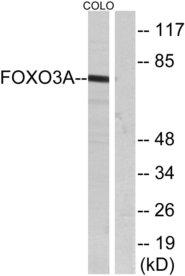
- Western blot analysis of lysates from COLO205 cells, treated with serum, using FKHRL1 Antibody. The lane on the right is blocked with the synthesized peptide.

- Western blot analysis of lysates from 1)MCF-7 , 2)COLO205, 3) HELA cells, (Green) primary antibody was diluted at 1:1000, 4°over night, secondary antibody(cat:RS23920)was diluted at 1:10000, 37° 1hour. (Red) Actin β Monoclonal Antibody(5B7) (cat:YM3028) antibody was diluted at 1:5000 as loading control, 4° over night,secondary antibody(cat:RS23710)was diluted at 1:10000, 37° 1hour.


