N-Cadherin (ABT-CDH2) mouse mAb
- Catalog No.:YM6590
- Applications:WB; IHC;ELISA
- Reactivity:Human;Mouse;(predicted: Rat)
- Target:
- N-Cadherin
- Fields:
- >>Cell adhesion molecules;>>Arrhythmogenic right ventricular cardiomyopathy
- Gene Name:
- CDH2 CDHN NCAD
- Protein Name:
- Cadherin-2 (CDw325) (Neural cadherin) (N-cadherin) (CD antigen CD325)
- Human Gene Id:
- 1000
- Human Swiss Prot No:
- P19022
- Immunogen:
- Synthesized peptide derived from human N-Cadherin AA range: 200-400
- Specificity:
- This antibody detects endogenous levels of human N-Cadherin. Heat-induced epitope retrieval (HIER) Citrate buffer of pH6.0 was highly recommended as antigen repair method in paraffin section
- Formulation:
- Liquid in PBS containing 50% glycerol, 0.5% BSA and 0.02% sodium azide.
- Source:
- Mouse, Monoclonal/IgG2b, Kappa
- Dilution:
- IHC 1:200-400,WB 1:500-2000, ELISA 1:5000-20000
- Purification:
- The antibody was affinity-purified from mouse ascites by affinity-chromatography using specific immunogen.
- Storage Stability:
- -15°C to -25°C/1 year(Do not lower than -25°C)
- Molecular Weight(Da):
- 100kD
- Background:
- This gene encodes a classical cadherin and member of the cadherin superfamily. Alternative splicing results in multiple transcript variants, at least one of which encodes a preproprotein is proteolytically processed to generate a calcium-dependent cell adhesion molecule and glycoprotein. This protein plays a role in the establishment of left-right asymmetry, development of the nervous system and the formation of cartilage and bone. [provided by RefSeq, Nov 2015],
- Function:
- function:Cadherins are calcium dependent cell adhesion proteins. They preferentially interact with themselves in a homophilic manner in connecting cells; cadherins may thus contribute to the sorting of heterogeneous cell types. CDH2 may be involved in neuronal recognition mechanism.,similarity:Contains 5 cadherin domains.,subunit:Interacts with CDCP1.,
- Subcellular Location:
- Cell membrane ; Single-pass type I membrane protein . Cell membrane, sarcolemma . Cell junction . Cell surface . Colocalizes with TMEM65 at the intercalated disk in cardiomyocytes. Colocalizes with OBSCN at the intercalated disk and at sarcolemma in cardiomyocytes. .
- Expression:
- Brain,Epithelium,Liver,
TP53I11 suppresses epithelial-mesenchymal transition and metastasis of breast cancer cells. BMB Reports Bmb Rep. 2019 Jun; 52(6): 379–384 WB Human MDA-MB-231 cell
- June 19-2018
- WESTERN IMMUNOBLOTTING PROTOCOL
- June 19-2018
- IMMUNOHISTOCHEMISTRY-PARAFFIN PROTOCOL
- June 19-2018
- IMMUNOFLUORESCENCE PROTOCOL
- September 08-2020
- FLOW-CYTOMEYRT-PROTOCOL
- May 20-2022
- Cell-Based ELISA│解您多样本WB检测之困扰
- July 13-2018
- CELL-BASED-ELISA-PROTOCOL-FOR-ACETYL-PROTEIN
- July 13-2018
- CELL-BASED-ELISA-PROTOCOL-FOR-PHOSPHO-PROTEIN
- July 13-2018
- Antibody-FAQs
- Products Images
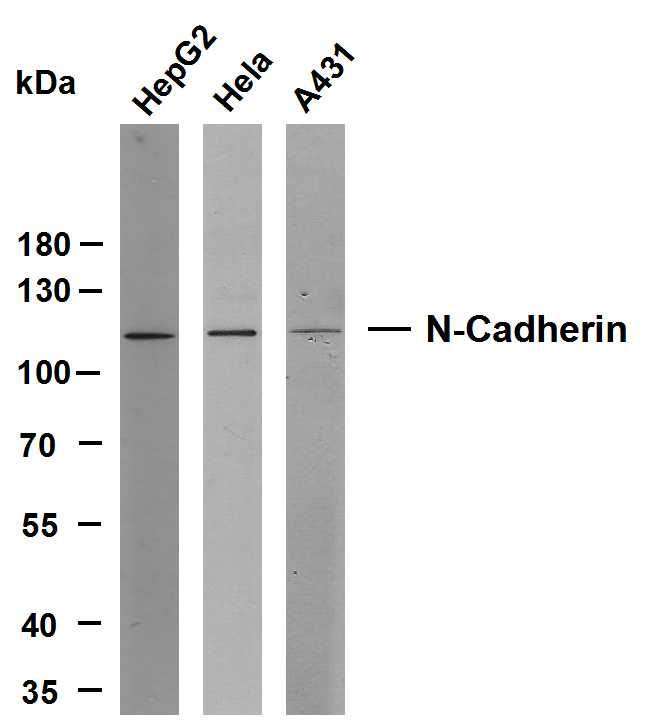
- Various whole cell lysates were separated by 8% SDS-PAGE, and the membrane was blotted with anti-N-Cadherin(ABT-CDH2) antibody. The HRP-conjugated Goat anti-Mouse IgG(H + L) antibody was used to detect the antibody. Lane 1: HepG2 Lane 2: Hela Lane 3: A431 Predicted band size: 100kDa Observed band size: 110kDa
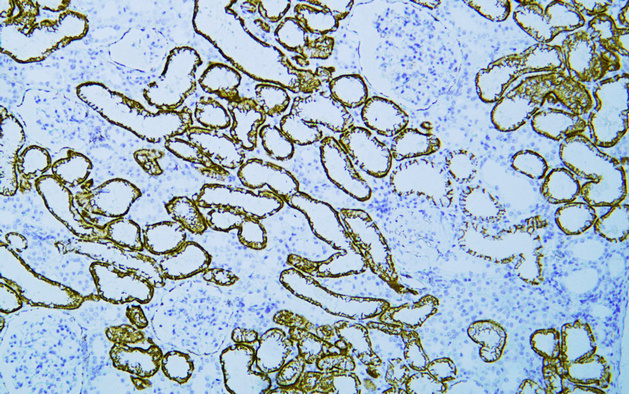
- Human Kidney tissue was stained with Anti-N-Cadherin (ABT-CDH2) Antibody
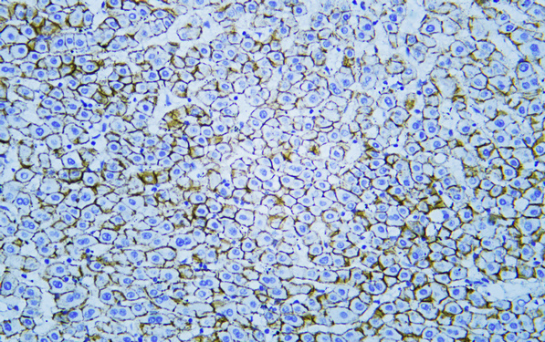
- Human liver tissue was stained with Anti-N-Cadherin (ABT-CDH2) Antibody

- Human liver tissue was stained with Anti-N-Cadherin (ABT-CDH2) Antibody
.jpg)
- Immunohistochemical analysis of paraffin-embedded Liver. 1, Antibody was diluted at 1:200(4° overnight). 2, Citrate buffer of pH6.0 was used for antigen retrieval. 3,Secondary antibody was diluted at 1:200(room temperature, 30min).
.jpg)
- Immunohistochemical analysis of paraffin-embedded Liver. 1, Antibody was diluted at 1:200(4° overnight). 2, Citrate buffer of pH6.0 was used for antigen retrieval. 3,Secondary antibody was diluted at 1:200(room temperature, 30min).
_wb.jpg)
- Western blot analysis of N-CadherinAntibody at 1:1000 dilution.
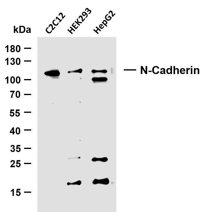
- Various whole cell lysates were separated by 15% SDS-PAGE, and the membrane was blotted with anti-N-Cadherin (ABT-CDH2) antibody. The HRP-conjugated Goat anti-Mouse IgG(H + L) antibody was used to detect the antibody. Lane 1: C2C12 Lane 2: HEK293 Lane 2: HepG2 Predicted band size: 100kDa Observed band size: 110kDa


