Oct1 Monoclonal Antibody(7G1)
- Catalog No.:YM3089
- Applications:WB;IHC;IF;
- Reactivity:Human
- Target:
- OCT1
- Fields:
- >>Herpes simplex virus 1 infection;>>Lipid and atherosclerosis
- Gene Name:
- POU2F1
- Protein Name:
- POU domain class 2 transcription factor
- Human Gene Id:
- 5451/5452
- Human Swiss Prot No:
- P14859/P09086
- Mouse Gene Id:
- 18986/18987
- Rat Gene Id:
- 171068
- Rat Swiss Prot No:
- P31503
- Immunogen:
- Synthetic Peptide of 42644
- Specificity:
- The antibody detects endogenous Oct1 protein.
- Formulation:
- PBS, pH 7.4, containing 0.5%BSA, 0.02% sodium azide as Preservative and 50% Glycerol.
- Source:
- Monoclonal, Mouse
- Dilution:
- WB 1:500-2000 IF 1:200 IHC 1:50-300
- Purification:
- The antibody was affinity-purified from mouse ascites by affinity-chromatography using specific immunogen.
- Storage Stability:
- -15°C to -25°C/1 year(Do not lower than -25°C)
- Observed Band(KD):
- 89kD
- Background:
- The OCT1 transcription factor was among the first identified members of the POU transcription factor family (summarized by Sturm et al., 1993 [PubMed 8314572]). Members of this family contain the POU domain, a 160-amino acid region necessary for DNA binding to the octameric sequence ATGCAAAT.[supplied by OMIM, Jul 2010],
- Function:
- function:Transcription factor that binds to the octamer motif (5'-ATTTGCAT-3') and activates the promoters of the genes for some small nuclear RNAs (snRNA) and of genes such as those for histone H2B and immunoglobulins. Modulates transcription transactivation by NR3C1, AR and PGR.,PTM:Phosphorylated by PRKDC.,similarity:Belongs to the POU transcription factor family. Class-2 subfamily.,similarity:Contains 1 homeobox DNA-binding domain.,similarity:Contains 1 POU-specific domain.,subunit:Interacts with NR3C1, AR, PGR and HCFC1.,tissue specificity:Ubiquitous. Isoform 2 is lymphocyte-specific.,
- Subcellular Location:
- Nucleus.
- Expression:
- Ubiquitous. Isoform 2 is lymphocyte-specific.
- June 19-2018
- WESTERN IMMUNOBLOTTING PROTOCOL
- June 19-2018
- IMMUNOHISTOCHEMISTRY-PARAFFIN PROTOCOL
- June 19-2018
- IMMUNOFLUORESCENCE PROTOCOL
- September 08-2020
- FLOW-CYTOMEYRT-PROTOCOL
- May 20-2022
- Cell-Based ELISA│解您多样本WB检测之困扰
- July 13-2018
- CELL-BASED-ELISA-PROTOCOL-FOR-ACETYL-PROTEIN
- July 13-2018
- CELL-BASED-ELISA-PROTOCOL-FOR-PHOSPHO-PROTEIN
- July 13-2018
- Antibody-FAQs
- Products Images
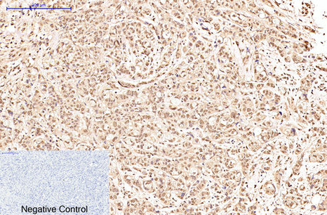
- Immunohistochemical analysis of paraffin-embedded Human-breast-cancer tissue. 1,Oct1 Monoclonal Antibody(7G1) was diluted at 1:200(4°C,overnight). 2, Sodium citrate pH 6.0 was used for antibody retrieval(>98°C,20min). 3,Secondary antibody was diluted at 1:200(room tempeRature, 30min). Negative control was used by secondary antibody only.
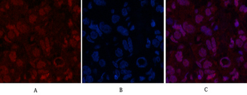
- Immunofluorescence analysis of Human-breast-cancer tissue. 1,Oct1 Monoclonal Antibody(7G1)(red) was diluted at 1:200(4°C,overnight). 2, Cy3 labled Secondary antibody was diluted at 1:300(room temperature, 50min).3, Picture B: DAPI(blue) 10min. Picture A:Target. Picture B: DAPI. Picture C: merge of A+B
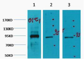
- Western blot analysis of 1) Hela, 2) Jurkat, 3) HepG2, diluted at 1:2000 cells nucleus extracted by Minute TM Cytoplasmic and Nuclear Fractionation kit (SC-003,Inventbiotech,MN,USA).


