Villin Polyclonal Antibody
- Catalog No.:YT5747
- Applications:WB;ELISA
- Reactivity:Human;Mouse;Rat
- Target:
- Villin
- Gene Name:
- VIL1 VIL
- Protein Name:
- Villin
- Human Gene Id:
- 7429
- Human Swiss Prot No:
- P09327
- Mouse Gene Id:
- 22349
- Mouse Swiss Prot No:
- Q62468
- Immunogen:
- Synthesized peptide derived from Villin at AA range: 601-650
- Specificity:
- Villin Polyclonal Antibody detects endogenous levels of Villin
- Formulation:
- Liquid in PBS containing 50% glycerol, 0.5% BSA and 0.02% sodium azide.
- Source:
- Polyclonal, Rabbit,IgG
- Dilution:
- WB 1:500-2000, ELISA 1:10000-20000
- Purification:
- The antibody was affinity-purified from rabbit antiserum by affinity-chromatography using epitope-specific immunogen.
- Concentration:
- 1 mg/ml
- Storage Stability:
- -15°C to -25°C/1 year(Do not lower than -25°C)
- Other Name:
- villin 1
- Observed Band(KD):
- 90kD
- Background:
- This gene encodes a member of a family of calcium-regulated actin-binding proteins. This protein represents a dominant part of the brush border cytoskeleton which functions in the capping, severing, and bundling of actin filaments. Two mRNAs of 2.7 kb and 3.5 kb have been observed; they result from utilization of alternate poly-adenylation signals present in the terminal exon. [provided by RefSeq, Jul 2008],
- Function:
- domain:Consists of a large core fragment, the N-terminal portion, and a small headpiece, the C-terminal portion. The headpiece binds F-actin strongly in both the presence and absence of calcium.,function:Ca(2+)-regulated actin-binding protein.,similarity:Belongs to the villin/gelsolin family.,similarity:Contains 1 HP (headpiece) domain.,similarity:Contains 6 gelsolin-like repeats.,subunit:Monomer.,tissue specificity:Major component of microvilli of intestinal epithelial cells and kidney proximal tubule cells.,
- Subcellular Location:
- Cytoplasm, cytoskeleton. Cell projection, lamellipodium. Cell projection, ruffle. Cell projection, microvillus. Cell projection, filopodium tip . Cell projection, filopodium . Relocalized in the tip of cellular protrusions and filipodial extensions upon infection with S.flexneri in primary intestinal epithelial cells (IEC) and in the tail-like structures forming the actin comets of S.flexneri. Redistributed to the leading edge of hepatocyte growth factor (HGF)-induced lamellipodia (By similarity). Rapidly redistributed to ruffles and lamellipodia structures in response to autotaxin, lysophosphatidic acid (LPA) and epidermal growth factor (EGF) treatment. .
- Expression:
- Specifically expressed in epithelial cells. Major component of microvilli of intestinal epithelial cells and kidney proximal tubule cells. Expressed in canalicular microvilli of hepatocytes (at protein level).
- June 19-2018
- WESTERN IMMUNOBLOTTING PROTOCOL
- June 19-2018
- IMMUNOHISTOCHEMISTRY-PARAFFIN PROTOCOL
- June 19-2018
- IMMUNOFLUORESCENCE PROTOCOL
- September 08-2020
- FLOW-CYTOMEYRT-PROTOCOL
- May 20-2022
- Cell-Based ELISA│解您多样本WB检测之困扰
- July 13-2018
- CELL-BASED-ELISA-PROTOCOL-FOR-ACETYL-PROTEIN
- July 13-2018
- CELL-BASED-ELISA-PROTOCOL-FOR-PHOSPHO-PROTEIN
- July 13-2018
- Antibody-FAQs
- Products Images
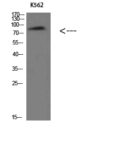
- Western Blot analysis of K562 cells using Villin Polyclonal Antibody diluted at 1:500. Secondary antibody(catalog#:RS0002) was diluted at 1:20000

- Immunohistochemical analysis of paraffin-embedded human-colon, antibody was diluted at 1:200


