TGFβ3 Polyclonal Antibody
- Catalog No.:YT4634
- Applications:WB;IHC;IF;ELISA
- Reactivity:Human;Mouse;Rat;Monkey
- Target:
- TGFB3
- Fields:
- >>MAPK signaling pathway;>>Cytokine-cytokine receptor interaction;>>FoxO signaling pathway;>>Cell cycle;>>Cellular senescence;>>TGF-beta signaling pathway;>>Hippo signaling pathway;>>AGE-RAGE signaling pathway in diabetic complications;>>Leishmaniasis;>>Chagas disease;>>Malaria;>>Toxoplasmosis;>>Amoebiasis;>>Tuberculosis;>>Hepatitis B;>>Human T-cell leukemia virus 1 infection;>>Pathways in cancer;>>Colorectal cancer;>>Renal cell carcinoma;>>Pancreatic cancer;>>Chronic myeloid leukemia;>>Hepatocellular carcinoma;>>Gastric cancer;>>Inflammatory bowel disease;>>Rheumatoid arthritis;>>Hypertrophic cardiomyopathy;>>Dilated cardiomyopathy;>>Diabetic cardiomyopathy
- Gene Name:
- TGFB3
- Protein Name:
- Transforming growth factor beta-3
- Human Gene Id:
- 7043
- Human Swiss Prot No:
- P10600
- Mouse Swiss Prot No:
- P17125
- Rat Gene Id:
- 25717
- Rat Swiss Prot No:
- Q07258
- Immunogen:
- The antiserum was produced against synthesized peptide derived from human TGF beta3. AA range:261-310
- Specificity:
- TGFβ3 Polyclonal Antibody detects endogenous levels of TGFβ3 protein.
- Formulation:
- Liquid in PBS containing 50% glycerol, 0.5% BSA and 0.02% sodium azide.
- Source:
- Polyclonal, Rabbit,IgG
- Dilution:
- WB 1:500 - 1:2000. IHC 1:100 - 1:300. ELISA: 1:10000.. IF 1:50-200
- Purification:
- The antibody was affinity-purified from rabbit antiserum by affinity-chromatography using epitope-specific immunogen.
- Concentration:
- 1 mg/ml
- Storage Stability:
- -15°C to -25°C/1 year(Do not lower than -25°C)
- Other Name:
- TGFB3;Transforming growth factor beta-3;TGF-beta-3
- Observed Band(KD):
- 13kD
- Background:
- This gene encodes a secreted ligand of the TGF-beta (transforming growth factor-beta) superfamily of proteins. Ligands of this family bind various TGF-beta receptors leading to recruitment and activation of SMAD family transcription factors that regulate gene expression. The encoded preproprotein is proteolytically processed to generate a latency-associated peptide (LAP) and a mature peptide, and is found in either a latent form composed of a mature peptide homodimer, a LAP homodimer, and a latent TGF-beta binding protein, or in an active form consisting solely of the mature peptide homodimer. The mature peptide may also form heterodimers with other TGF-beta family members. This protein is involved in embryogenesis and cell differentiation, and may play a role in wound healing. Mutations in this gene are a cause of aortic aneurysms and dissections, as well as familial arrhythmogenic
- Function:
- disease:Defects in TGFB3 are a cause of familial arrhythmogenic right ventricular dysplasia 1 (ARVD1) [MIM:107970]; also known as arrhythmogenic right ventricular cardiomyopathy 1 (ARVC1). ARVD is an autosomal dominant disease characterized by partial degeneration of the myocardium of the right ventricle, electrical instability, and sudden death. It is clinically defined by electrocardiographic and angiographic criteria; pathologic findings, replacement of ventricular myocardium with fatty and fibrous elements, preferentially involve the right ventricular free wall.,function:Involved in embryogenesis and cell differentiation.,online information:TGF beta-3 entry,similarity:Belongs to the TGF-beta family.,subunit:Homodimer; disulfide-linked.,
- Subcellular Location:
- [Latency-associated peptide]: Secreted, extracellular space, extracellular matrix .; [Transforming growth factor beta-3]: Secreted .
- Expression:
- Amygdala,Esophageal,Placenta,Prostate,
- June 19-2018
- WESTERN IMMUNOBLOTTING PROTOCOL
- June 19-2018
- IMMUNOHISTOCHEMISTRY-PARAFFIN PROTOCOL
- June 19-2018
- IMMUNOFLUORESCENCE PROTOCOL
- September 08-2020
- FLOW-CYTOMEYRT-PROTOCOL
- May 20-2022
- Cell-Based ELISA│解您多样本WB检测之困扰
- July 13-2018
- CELL-BASED-ELISA-PROTOCOL-FOR-ACETYL-PROTEIN
- July 13-2018
- CELL-BASED-ELISA-PROTOCOL-FOR-PHOSPHO-PROTEIN
- July 13-2018
- Antibody-FAQs
- Products Images
- Western Blot analysis of various cells using TGFβ3 Polyclonal Antibody

- Immunohistochemistry analysis of paraffin-embedded human breast carcinoma tissue, using TGF beta3 Antibody. The picture on the right is blocked with the synthesized peptide.

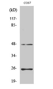
- Western blot analysis of lysates from COS7 cells, using TGF beta3 Antibody. The lane on the right is blocked with the synthesized peptide.