Tak1 Polyclonal Antibody
- Catalog No.:YT4536
- Applications:IF;WB;IHC;ELISA
- Reactivity:Human;Mouse;Rat
- Target:
- Tak1
- Fields:
- >>MAPK signaling pathway;>>NF-kappa B signaling pathway;>>Autophagy - animal;>>AMPK signaling pathway;>>Wnt signaling pathway;>>Osteoclast differentiation;>>Adherens junction;>>Neutrophil extracellular trap formation;>>Toll-like receptor signaling pathway;>>NOD-like receptor signaling pathway;>>RIG-I-like receptor signaling pathway;>>IL-17 signaling pathway;>>T cell receptor signaling pathway;>>TNF signaling pathway;>>Alcoholic liver disease;>>Pathogenic Escherichia coli infection;>>Shigellosis;>>Salmonella infection;>>Yersinia infection;>>Leishmaniasis;>>Toxoplasmosis;>>Hepatitis B;>>Measles;>>Herpes simplex virus 1 infection;>>Epstein-Barr virus infection;>>Human immunodeficiency virus 1 infection;>>Coronavirus disease - COVID-19;>>Lipid and atherosclerosis;>>Fluid shear stress and atherosclerosis
- Gene Name:
- MAP3K7
- Protein Name:
- Mitogen-activated protein kinase kinase kinase 7
- Human Gene Id:
- 6885
- Human Swiss Prot No:
- O43318
- Mouse Gene Id:
- 26409
- Mouse Swiss Prot No:
- Q62073
- Rat Gene Id:
- 1.00911e+008
- Rat Swiss Prot No:
- P0C8E4
- Immunogen:
- The antiserum was produced against synthesized peptide derived from human MAP3K7. AA range:161-210
- Specificity:
- Tak1 Polyclonal Antibody detects endogenous levels of Tak1 protein.
- Formulation:
- Liquid in PBS containing 50% glycerol, 0.5% BSA and 0.02% sodium azide.
- Source:
- Polyclonal, Rabbit,IgG
- Dilution:
- IF 1:50-200 WB 1:500 - 1:2000. IHC 1:100 - 1:300. ELISA: 1:40000. Not yet tested in other applications.
- Purification:
- The antibody was affinity-purified from rabbit antiserum by affinity-chromatography using epitope-specific immunogen.
- Concentration:
- 1 mg/ml
- Storage Stability:
- -15°C to -25°C/1 year(Do not lower than -25°C)
- Other Name:
- MAP3K7;TAK1;Mitogen-activated protein kinase kinase kinase 7;Transforming growth factor-beta-activated kinase 1;TGF-beta-activated kinase 1
- Observed Band(KD):
- 77kD
- Background:
- The protein encoded by this gene is a member of the serine/threonine protein kinase family. This kinase mediates the signaling transduction induced by TGF beta and morphogenetic protein (BMP), and controls a variety of cell functions including transcription regulation and apoptosis. In response to IL-1, this protein forms a kinase complex including TRAF6, MAP3K7P1/TAB1 and MAP3K7P2/TAB2; this complex is required for the activation of nuclear factor kappa B. This kinase can also activate MAPK8/JNK, MAP2K4/MKK4, and thus plays a role in the cell response to environmental stresses. Four alternatively spliced transcript variants encoding distinct isoforms have been reported. [provided by RefSeq, Jul 2008],
- Function:
- catalytic activity:ATP + a protein = ADP + a phosphoprotein.,cofactor:Magnesium.,function:Component of a protein kinase signal transduction cascade. Mediator of TGF-beta signal transduction. Stimulates NF-kappa-B activation and the p38 MAPK pathway.,PTM:Association with MAP3K7IP1 promotes autophosphorylation and subsequent activation. Dephosphorylation at Thr-187 by PP2A and PPP6C leads to inactivation.,similarity:Belongs to the protein kinase superfamily.,similarity:Belongs to the protein kinase superfamily. STE Ser/Thr protein kinase family. MAP kinase kinase kinase subfamily.,similarity:Contains 1 protein kinase domain.,subunit:Binds both upstream activators and downstream substrates in multimolecular complexes. Interacts with MAP3K7IP1 and MAP3K7IP2. Interacts with PPM1L. Interaction with PP2A and PPP6C leads to its' repressed activity.,
- Subcellular Location:
- Cytoplasm . Cell membrane ; Peripheral membrane protein ; Cytoplasmic side . Although the majority of MAP3K7/TAK1 is found in the cytosol, when complexed with TAB1/MAP3K7IP1 and TAB2/MAP3K7IP2, it is also localized at the cell membrane.
- Expression:
- Isoform 1A is the most abundant in ovary, skeletal muscle, spleen and blood mononuclear cells. Isoform 1B is highly expressed in brain, kidney and small intestine. Isoform 1C is the major form in prostate. Isoform 1D is the less abundant form.
RNA-induced liquid phase separation of SARS-CoV-2 nucleocapsid protein facilitates NF-κB hyper-activation and inflammation. Signal Transduction and Targeted Therapy Signal Transduct Tar. 2021 Apr;6(1):1-13 IF,WB Human HEK293T cell
Exercise ameliorates insulin resistance via regulating TGFβ‐activated kinase 1 (TAK1)‐mediated insulin signaling in liver of high‐fat diet‐induced obese rats. JOURNAL OF CELLULAR PHYSIOLOGY 2018 Oct 26 WB Rat 1:1000 liver tissues
Exercise ameliorates insulin resistance via regulating TGFβ‐activated kinase 1 (TAK1)‐mediated insulin signaling in liver of high‐fat diet‐induced obese rats. JOURNAL OF CELLULAR PHYSIOLOGY 2018 Oct 26 WB Rat 1:1000 liver tissues
Exercise ameliorates insulin resistance via regulating TGFβ‐activated kinase 1 (TAK1)‐mediated insulin signaling in liver of high‐fat diet‐induced obese rats. JOURNAL OF CELLULAR PHYSIOLOGY 2018 Oct 26 WB Rat 1:1000 liver tissues
Alpha-Momorcharin Inhibits Proinflammatory Cytokine Expression by M1 Macrophages but Not Anti-Inflammatory Cytokine Expression by M2 Macrophages Journal of Inflammation Research Fubing Shen WB Human
NLRC5 Deficiency Reduces LPS-Induced Microglial Activation via Inhibition of NF-κB Signaling and Ameliorates Mice’s Depressive-like Behavior. INTERNATIONAL JOURNAL OF MOLECULAR SCIENCES Jianqiong Zhang WB Mouse 1:1000 BV2 cell
Upregulation of TRIM16 mitigates doxorubicin-induced cardiotoxicity by modulating TAK1 and YAP/Nrf2 pathways in mice BIOCHEMICAL PHARMACOLOGY Xinyu Guo WB,IP Mouse,Rat 1:1000 heart tissue Neonatal rat cardiomyocytes (NRCMs)
- June 19-2018
- WESTERN IMMUNOBLOTTING PROTOCOL
- June 19-2018
- IMMUNOHISTOCHEMISTRY-PARAFFIN PROTOCOL
- June 19-2018
- IMMUNOFLUORESCENCE PROTOCOL
- September 08-2020
- FLOW-CYTOMEYRT-PROTOCOL
- May 20-2022
- Cell-Based ELISA│解您多样本WB检测之困扰
- July 13-2018
- CELL-BASED-ELISA-PROTOCOL-FOR-ACETYL-PROTEIN
- July 13-2018
- CELL-BASED-ELISA-PROTOCOL-FOR-PHOSPHO-PROTEIN
- July 13-2018
- Antibody-FAQs
- Products Images
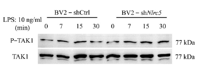
- NLRC5 Deficiency Reduces LPS-Induced Microglial Activation via Inhibition of NF-κB Signaling and Ameliorates Mice’s Depressive-like Behavior. INTERNATIONAL JOURNAL OF MOLECULAR SCIENCES Jianqiong Zhang WB Mouse 1:500 BV2 cell
.jpg)
- Wu, Y., Ma, L., Cai, S. et al. RNA-induced liquid phase separation of SARS-CoV-2 nucleocapsid protein facilitates NF-κB hyper-activation and inflammation. Sig Transduct Target Ther 6, 167 (2021)

- Immunofluorescence analysis of Hela cell. 1,Tak1 Polyclonal Antibody(red) was diluted at 1:200(4° overnight). CK7 Monoclonal Antibody(12D7)(green) was diluted at 1:200(4° overnight). 2, Goat Anti Rabbit Alexa Fluor 594 Catalog:RS3611 was diluted at 1:1000(room temperature, 50min). Goat Anti Mouse Alexa Fluor 488 Catalog:RS3208 was diluted at 1:1000(room temperature, 50min).

- Immunofluorescence analysis of human-stomach tissue. 1,Tak1 Polyclonal Antibody(red) was diluted at 1:200(4°C,overnight). 2, Cy3 labled Secondary antibody was diluted at 1:300(room temperature, 50min).3, Picture B: DAPI(blue) 10min. Picture A:Target. Picture B: DAPI. Picture C: merge of A+B

- Immunofluorescence analysis of human-stomach tissue. 1,Tak1 Polyclonal Antibody(red) was diluted at 1:200(4°C,overnight). 2, Cy3 labled Secondary antibody was diluted at 1:300(room temperature, 50min).3, Picture B: DAPI(blue) 10min. Picture A:Target. Picture B: DAPI. Picture C: merge of A+B

- Immunofluorescence analysis of rat-kidney tissue. 1,Tak1 Polyclonal Antibody(red) was diluted at 1:200(4°C,overnight). 2, Cy3 labled Secondary antibody was diluted at 1:300(room temperature, 50min).3, Picture B: DAPI(blue) 10min. Picture A:Target. Picture B: DAPI. Picture C: merge of A+B

- Immunofluorescence analysis of rat-kidney tissue. 1,Tak1 Polyclonal Antibody(red) was diluted at 1:200(4°C,overnight). 2, Cy3 labled Secondary antibody was diluted at 1:300(room temperature, 50min).3, Picture B: DAPI(blue) 10min. Picture A:Target. Picture B: DAPI. Picture C: merge of A+B

- Immunofluorescence analysis of mouse-kidney tissue. 1,Tak1 Polyclonal Antibody(red) was diluted at 1:200(4°C,overnight). 2, Cy3 labled Secondary antibody was diluted at 1:300(room temperature, 50min).3, Picture B: DAPI(blue) 10min. Picture A:Target. Picture B: DAPI. Picture C: merge of A+B

- Immunofluorescence analysis of mouse-kidney tissue. 1,Tak1 Polyclonal Antibody(red) was diluted at 1:200(4°C,overnight). 2, Cy3 labled Secondary antibody was diluted at 1:300(room temperature, 50min).3, Picture B: DAPI(blue) 10min. Picture A:Target. Picture B: DAPI. Picture C: merge of A+B

- Immunohistochemical analysis of paraffin-embedded Human-stomach tissue. 1,Tak1 Polyclonal Antibody was diluted at 1:200(4°C,overnight). 2, Sodium citrate pH 6.0 was used for antibody retrieval(>98°C,20min). 3,Secondary antibody was diluted at 1:200(room tempeRature, 30min). Negative control was used by secondary antibody only.

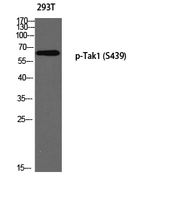
- Western Blot analysis of various cells using Tak1 Polyclonal Antibody diluted at 1:1000

- Immunohistochemistry analysis of paraffin-embedded human breast carcinoma tissue, using MAP3K7 Antibody. The picture on the right is blocked with the synthesized peptide.

- Western blot analysis of lysates from Jurkat cells, treated with heat shock, using MAP3K7 Antibody. The lane on the right is blocked with the synthesized peptide.