PPAR-γ Polyclonal Antibody
- Catalog No.:YT3836
- Applications:IF;WB;IHC;ELISA
- Reactivity:Human;Mouse;Rat
- Target:
- PPAR-γ
- Fields:
- >>PPAR signaling pathway;>>AMPK signaling pathway;>>Longevity regulating pathway;>>Osteoclast differentiation;>>Thermogenesis;>>Non-alcoholic fatty liver disease;>>Huntington disease;>>Pathways in cancer;>>Transcriptional misregulation in cancer;>>Thyroid cancer;>>Lipid and atherosclerosis
- Gene Name:
- PPARG
- Protein Name:
- Peroxisome proliferator-activated receptor gamma
- Human Gene Id:
- 5468
- Human Swiss Prot No:
- P37231
- Mouse Gene Id:
- 19016
- Mouse Swiss Prot No:
- P37238
- Rat Gene Id:
- 25664
- Rat Swiss Prot No:
- O88275
- Immunogen:
- The antiserum was produced against synthesized peptide derived from human PPAR-gamma. AA range:78-127
- Specificity:
- PPAR-γ Polyclonal Antibody detects endogenous levels of PPAR-γ protein.
- Formulation:
- Liquid in PBS containing 50% glycerol, 0.5% BSA and 0.02% sodium azide.
- Source:
- Polyclonal, Rabbit,IgG
- Dilution:
- IF 1:50-200 WB 1:500 - 1:2000. IHC 1:100 - 1:300. ELISA: 1:10000. Not yet tested in other applications.
- Purification:
- The antibody was affinity-purified from rabbit antiserum by affinity-chromatography using epitope-specific immunogen.
- Concentration:
- 1 mg/ml
- Storage Stability:
- -15°C to -25°C/1 year(Do not lower than -25°C)
- Other Name:
- PPARG;NR1C3;Peroxisome proliferator-activated receptor gamma;PPAR-gamma;Nuclear receptor subfamily 1 group C member 3
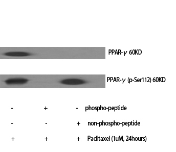
- Observed Band(KD):
- 57kD
- Background:
- peroxisome proliferator activated receptor gamma(PPARG) Homo sapiens This gene encodes a member of the peroxisome proliferator-activated receptor (PPAR) subfamily of nuclear receptors. PPARs form heterodimers with retinoid X receptors (RXRs) and these heterodimers regulate transcription of various genes. Three subtypes of PPARs are known: PPAR-alpha, PPAR-delta, and PPAR-gamma. The protein encoded by this gene is PPAR-gamma and is a regulator of adipocyte differentiation. Additionally, PPAR-gamma has been implicated in the pathology of numerous diseases including obesity, diabetes, atherosclerosis and cancer. Alternatively spliced transcript variants that encode different isoforms have been described. [provided by RefSeq, Jul 2008],
- Function:
- alternative products:Additional isoforms seem to exist,disease:Defects in PPARG are the cause of familial partial lipodystrophy type 3 (FPLD3) [MIM:604367]. Familial partial lipodystrophies (FPLD) are a heterogeneous group of genetic disorders characterized by marked loss of subcutaneous (sc) fat from the extremities. Affected individuals show an increased preponderance of insulin resistance, diabetes mellitus and dyslipidemia.,disease:Defects in PPARG can lead to type 2 insulin-resistant diabetes and hyptertension.,disease:Defects in PPARG may be associated with colon cancer.,disease:Defects in PPARG may be associated with susceptibility to obesity [MIM:601665].,disease:Variation in PPARG is associated with carotid intimal medial thickness 1 (CIMT1) [MIM:609338]. CIMT is a measure of atherosclerosis that is independently associated with traditional atherosclerotic cardiovascular disease
- Subcellular Location:
- Nucleus. Cytoplasm. Redistributed from the nucleus to the cytosol through a MAP2K1/MEK1-dependent manner. NOCT enhances its nuclear translocation.
- Expression:
- Highest expression in adipose tissue. Lower in skeletal muscle, spleen, heart and liver. Also detectable in placenta, lung and ovary.
The tibetan medicine Zuozhu-Daxi can prevent Helicobacter pylori induced-gastric mucosa inflammation by inhibiting lipid metabolism
Metformin Attenuates Cardiac Hypertrophy Via the HIF-1α/PPAR-γ Signaling Pathway in High-Fat Diet Rats WB Rat left ventricle/H9c2 cells
1,8-Cineole Ameliorates LPS-Induced Vascular Endothelium Dysfunction in Mice via PPAR-γ Dependent Regulation of NF-κB. Frontiers in Pharmacology Front Pharmacol. 2019 Mar;0:178 WB Mouse 1:1000 Thoracic aorta tissue
Taxifolin inhibits keratinocyte proliferation and ameliorates imiquimod-induced psoriasis-like mouse model via regulating cytoplasmic phospholipase A2 and PPAR-γ pathway. INTERNATIONAL IMMUNOPHARMACOLOGY Int Immunopharmacol. 2021 Oct;99:107900 WB Mouse 1:1000 Skin
1, 8-Cineole ameliorates LPS-induced vascular endothelium dysfunction in mice via PPAR-γ dependent regulation of NF-κB." Frontiers in Pharmacology 10 (2019): 178.
Bai, Yi-Hua, et al. "Acteoside ameliorates diabetic kidney disease via regulating the activation of the PPARγ/β-catenin pathway." SCIENCEASIA 47.3 (2021): 293-+.
Taxifolin inhibits keratinocyte proliferation and ameliorates imiquimod-induced psoriasis-like mouse model via regulating cytoplasmic phospholipase A2 and PPAR-γ pathway. INTERNATIONAL IMMUNOPHARMACOLOGY Int Immunopharmacol. 2021 Oct;99:107900 WB Mouse 1:1000 Skin
A novel multifunctional mitochondrion-targeting NIR fluorophore probe inhibits tumour proliferation and metastasis through the PPARγ/ROS/β-catenin pathway. Changzhen Sun WB,IHC Mouse,Human B16-F10 cell-Xenograft A375 cell,B16–F10 cell
IRE1α-JNK axis activation contributes to intracellular lipid accumulation in calf hepatocytes JOURNAL OF DAIRY SCIENCE Wenwen Gao WB Calf 1:500 hepatocytes
Integrated investigation and experimental validation of PPARG as an oncogenic driver: implications for prognostic assessment and therapeutic targeting in hepatocellular carcinoma Frontiers in Pharmacology Yunsheng Ran WB Human LM3 cell,Huh7 cell,HLF cell,Hep G2 cell,LO2 cell,L68 cell
Perfluorooctane sulfonate causes pyroptosis and lipid metabolism disorders through ROS-mediated NLRP3 inflammasome activation in grass carp hepatocyte AQUATIC TOXICOLOGY Bendong Shi WB Fish L8824 cell
- June 19-2018
- WESTERN IMMUNOBLOTTING PROTOCOL
- June 19-2018
- IMMUNOHISTOCHEMISTRY-PARAFFIN PROTOCOL
- June 19-2018
- IMMUNOFLUORESCENCE PROTOCOL
- September 08-2020
- FLOW-CYTOMEYRT-PROTOCOL
- May 20-2022
- Cell-Based ELISA│解您多样本WB检测之困扰
- July 13-2018
- CELL-BASED-ELISA-PROTOCOL-FOR-ACETYL-PROTEIN
- July 13-2018
- CELL-BASED-ELISA-PROTOCOL-FOR-PHOSPHO-PROTEIN
- July 13-2018
- Antibody-FAQs
- Products Images

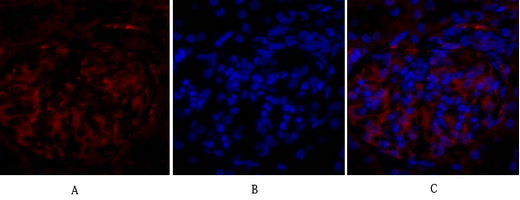
- Immunofluorescence analysis of rat-lung tissue. 1,PPAR-γ Polyclonal Antibody(red) was diluted at 1:200(4°C,overnight). 2, Cy3 labled Secondary antibody was diluted at 1:300(room temperature, 50min).3, Picture B: DAPI(blue) 10min. Picture A:Target. Picture B: DAPI. Picture C: merge of A+B
- Immunofluorescence analysis of rat-kidney tissue. 1,PPAR-γ Polyclonal Antibody(red) was diluted at 1:200(4°C,overnight). 2, Cy3 labled Secondary antibody was diluted at 1:300(room temperature, 50min).3, Picture B: DAPI(blue) 10min. Picture A:Target. Picture B: DAPI. Picture C: merge of A+B
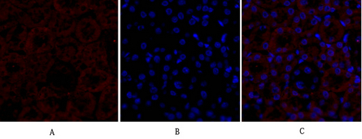
- Immunofluorescence analysis of mouse-kidney tissue. 1,PPAR-γ Polyclonal Antibody(red) was diluted at 1:200(4°C,overnight). 2, Cy3 labled Secondary antibody was diluted at 1:300(room temperature, 50min).3, Picture B: DAPI(blue) 10min. Picture A:Target. Picture B: DAPI. Picture C: merge of A+B
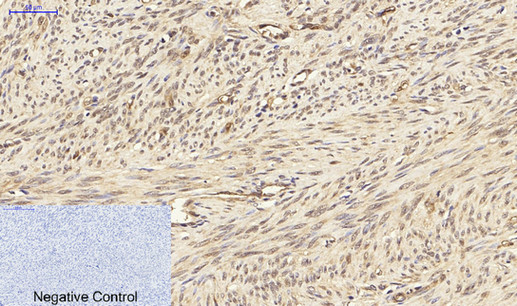
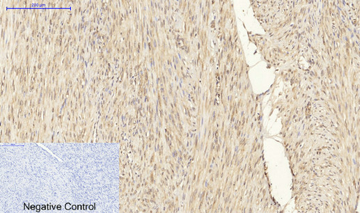
- Immunohistochemical analysis of paraffin-embedded Human-uterus tissue. 1,PPAR-γ Polyclonal Antibody was diluted at 1:200(4°C,overnight). 2, Sodium citrate pH 6.0 was used for antibody retrieval(>98°C,20min). 3,Secondary antibody was diluted at 1:200(room tempeRature, 30min). Negative control was used by secondary antibody only.
- Immunohistochemical analysis of paraffin-embedded Human-uterus-cancer tissue. 1,PPAR-γ Polyclonal Antibody was diluted at 1:200(4°C,overnight). 2, Sodium citrate pH 6.0 was used for antibody retrieval(>98°C,20min). 3,Secondary antibody was diluted at 1:200(room tempeRature, 30min). Negative control was used by secondary antibody only.
- Immunohistochemical analysis of paraffin-embedded Human-Tonsil tissue. 1,PPAR-γ Polyclonal Antibody was diluted at 1:200(4°C,overnight). 2, Sodium citrate pH 6.0 was used for antibody retrieval(>98°C,20min). 3,Secondary antibody was diluted at 1:200(room tempeRature, 30min). Negative control was used by secondary antibody only.
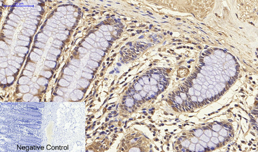
- Immunohistochemical analysis of paraffin-embedded Human-colon tissue. 1,PPAR-γ Polyclonal Antibody was diluted at 1:200(4°C,overnight). 2, Sodium citrate pH 6.0 was used for antibody retrieval(>98°C,20min). 3,Secondary antibody was diluted at 1:200(room tempeRature, 30min). Negative control was used by secondary antibody only.
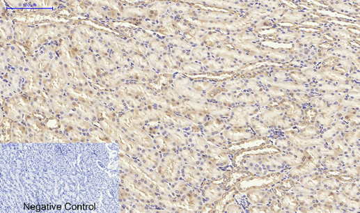
- Immunohistochemical analysis of paraffin-embedded Rat-kidney tissue. 1,PPAR-γ Polyclonal Antibody was diluted at 1:200(4°C,overnight). 2, Sodium citrate pH 6.0 was used for antibody retrieval(>98°C,20min). 3,Secondary antibody was diluted at 1:200(room tempeRature, 30min). Negative control was used by secondary antibody only.
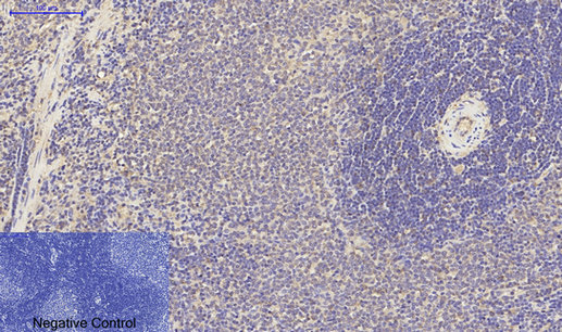
- Immunohistochemical analysis of paraffin-embedded Rat-spleen tissue. 1,PPAR-γ Polyclonal Antibody was diluted at 1:200(4°C,overnight). 2, Sodium citrate pH 6.0 was used for antibody retrieval(>98°C,20min). 3,Secondary antibody was diluted at 1:200(room tempeRature, 30min). Negative control was used by secondary antibody only.
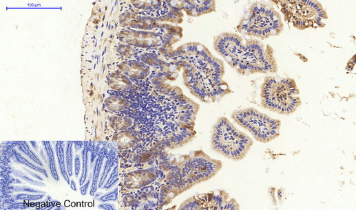
- Immunohistochemical analysis of paraffin-embedded Mouse-colon tissue. 1,PPAR-γ Polyclonal Antibody was diluted at 1:200(4°C,overnight). 2, Sodium citrate pH 6.0 was used for antibody retrieval(>98°C,20min). 3,Secondary antibody was diluted at 1:200(room tempeRature, 30min). Negative control was used by secondary antibody only.
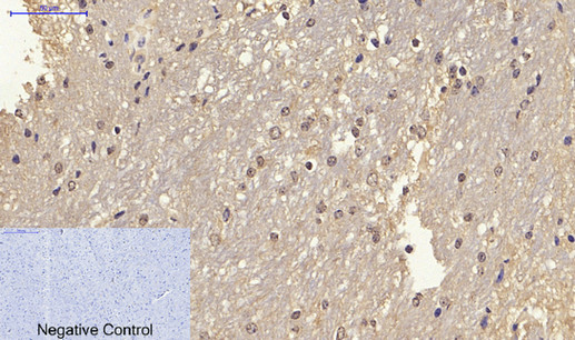
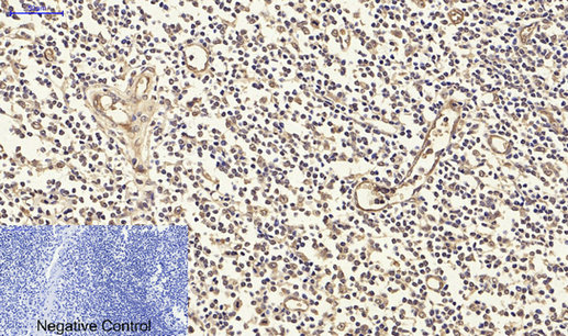
- Immunohistochemical analysis of paraffin-embedded Mouse-brain tissue. 1,PPAR-γ Polyclonal Antibody was diluted at 1:200(4°C,overnight). 2, Sodium citrate pH 6.0 was used for antibody retrieval(>98°C,20min). 3,Secondary antibody was diluted at 1:200(room tempeRature, 30min). Negative control was used by secondary antibody only.
- Western Blot analysis of various cells using PPAR-γ Polyclonal Antibody diluted at 1:1000
.jpg)
- Western Blot analysis of HuvEc cells using PPAR-γ Polyclonal Antibody diluted at 1:1000