CD83 Polyclonal Antibody
- Catalog No.:YT0779
- Applications:WB;IHC;IF;ELISA
- Reactivity:Human;Rat;Mouse;
- Target:
- CD83
- Gene Name:
- CD83
- Protein Name:
- CD83 antigen
- Human Gene Id:
- 9308
- Human Swiss Prot No:
- Q01151
- Mouse Swiss Prot No:
- O88324
- Immunogen:
- The antiserum was produced against synthesized peptide derived from human CD83. AA range:101-150
- Specificity:
- CD83 Polyclonal Antibody detects endogenous levels of CD83 protein.
- Formulation:
- Liquid in PBS containing 50% glycerol, 0.5% BSA and 0.02% sodium azide.
- Source:
- Polyclonal, Rabbit,IgG
- Dilution:
- WB 1:500 - 1:2000. IHC 1:100 - 1:300. ELISA: 1:10000.. IF 1:50-200
- Purification:
- The antibody was affinity-purified from rabbit antiserum by affinity-chromatography using epitope-specific immunogen.
- Concentration:
- 1 mg/ml
- Storage Stability:
- -15°C to -25°C/1 year(Do not lower than -25°C)
- Other Name:
- CD83;CD83 antigen;hCD83;B-cell activation protein;Cell surface protein HB15;CD antigen CD83
- Observed Band(KD):
- 23kD
- Background:
- The protein encoded by this gene is a single-pass type I membrane protein and member of the immunoglobulin superfamily of receptors. The encoded protein may be involved in the regulation of antigen presentation. A soluble form of this protein can bind to dendritic cells and inhibit their maturation. Three transcript variants encoding different isoforms have been found for this gene. [provided by RefSeq, Oct 2011],
- Function:
- function:May play a significant role in antigen presentation or the cellular interactions that follow lymphocyte activation.,online information:CD83 antigen,similarity:Contains 1 Ig-like V-type (immunoglobulin-like) domain.,subunit:Monomer.,tissue specificity:Expressed by activated lymphocytes, Langerhans cells and interdigitating reticulum cells.,
- Subcellular Location:
- Membrane; Single-pass type I membrane protein.
- Expression:
- Expressed by activated lymphocytes, Langerhans cells and interdigitating reticulum cells.
- June 19-2018
- WESTERN IMMUNOBLOTTING PROTOCOL
- June 19-2018
- IMMUNOHISTOCHEMISTRY-PARAFFIN PROTOCOL
- June 19-2018
- IMMUNOFLUORESCENCE PROTOCOL
- September 08-2020
- FLOW-CYTOMEYRT-PROTOCOL
- May 20-2022
- Cell-Based ELISA│解您多样本WB检测之困扰
- July 13-2018
- CELL-BASED-ELISA-PROTOCOL-FOR-ACETYL-PROTEIN
- July 13-2018
- CELL-BASED-ELISA-PROTOCOL-FOR-PHOSPHO-PROTEIN
- July 13-2018
- Antibody-FAQs
- Products Images
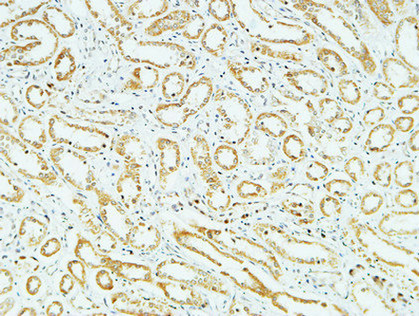
- Immunohistochemical analysis of paraffin-embedded Human kidney. 1, Antibody was diluted at 1:400(4° overnight). 2, High-pressure and temperature EDTA, pH8.0 was used for antigen retrieval. 3,Secondary antibody was diluted at 1:200(room temperature, 30min).
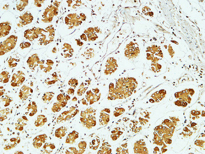
- Immunohistochemical analysis of paraffin-embedded Human stomach. 1, Antibody was diluted at 1:400(4° overnight). 2, High-pressure and temperature EDTA, pH8.0 was used for antigen retrieval. 3,Secondary antibody was diluted at 1:200(room temperature, 30min).

- Immunohistochemistry analysis of paraffin-embedded human lung carcinoma, using CD83 Antibody. The picture on the right is blocked with the synthesized peptide.
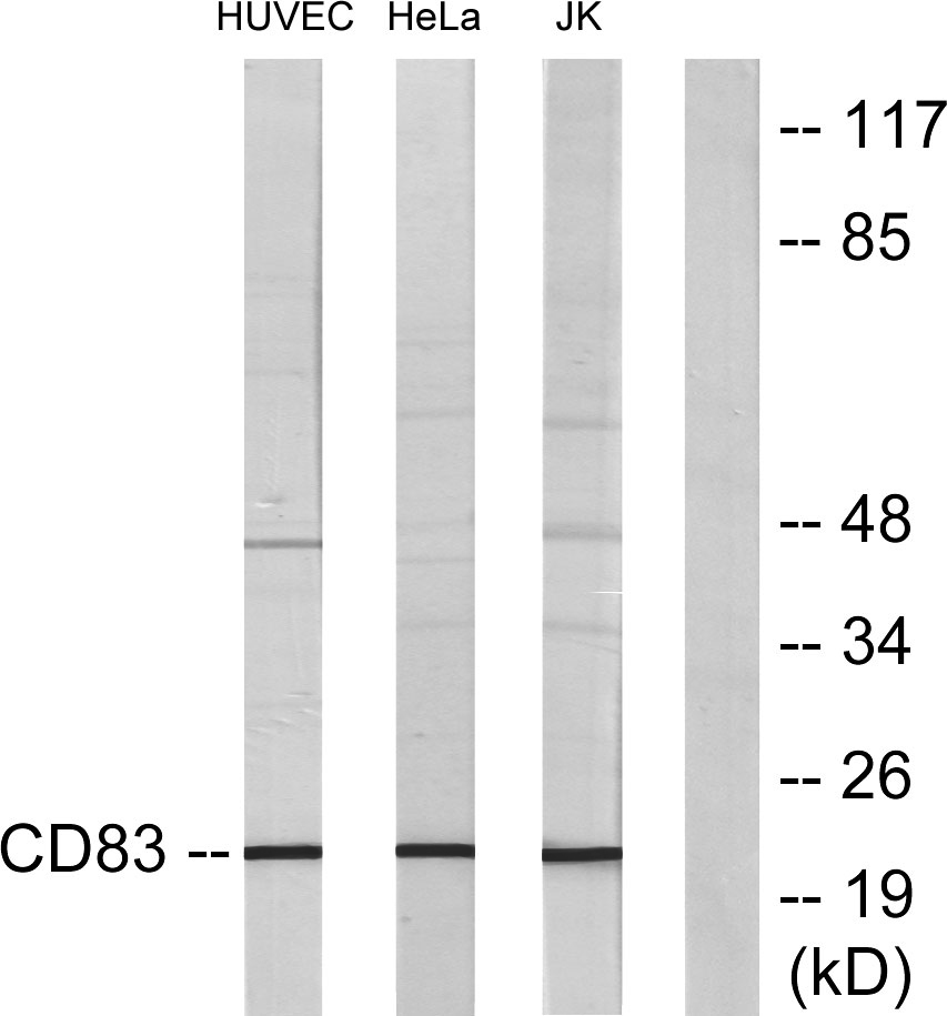
- Western blot analysis of lysates from HepG2 cells, HUVEC cells, HeLa cells, and Jurkat cells, using CD83 Antibody. The lane on the right is blocked with the synthesized peptide.


