14-3-3 σ Polyclonal Antibody
- Catalog No.:YT0012
- Applications:WB;IHC;IF;ELISA
- Reactivity:Human;Mouse
- Target:
- 14-3-3σ
- Fields:
- >>Cell cycle;>>p53 signaling pathway;>>Aldosterone-regulated sodium reabsorption
- Gene Name:
- SFN
- Protein Name:
- 14-3-3 protein sigma
- Human Gene Id:
- 2810
- Human Swiss Prot No:
- P31947
- Mouse Gene Id:
- 55948
- Mouse Swiss Prot No:
- O70456
- Immunogen:
- The antiserum was produced against synthesized peptide derived from human SFN. AA range:41-90
- Specificity:
- 14-3-3 σ Polyclonal Antibody detects endogenous levels of 14-3-3 σ protein.
- Formulation:
- Liquid in PBS containing 50% glycerol, 0.5% BSA and 0.02% sodium azide.
- Source:
- Polyclonal, Rabbit,IgG
- Dilution:
- IHC: 100-300.WB 1:500 - 1:2000. ELISA: 1:20000.. IF 1:50-200
- Purification:
- The antibody was affinity-purified from rabbit antiserum by affinity-chromatography using epitope-specific immunogen.
- Concentration:
- 1 mg/ml
- Storage Stability:
- -15°C to -25°C/1 year(Do not lower than -25°C)
- Other Name:
- SFN;HME1;14-3-3 protein sigma;Epithelial cell marker protein 1;Stratifin
- Observed Band(KD):
- 30kD
- Background:
- function:Adapter protein implicated in the regulation of a large spectrum of both general and specialized signaling pathway. Binds to a large number of partners, usually by recognition of a phosphoserine or phosphothreonine motif. Binding generally results in the modulation of the activity of the binding partner. When bound to KRT17, regulates protein synthesis and epithelial cell growth by stimulating Akt/mTOR pathway.,function:p53-regulated inhibitor of G2/M progression.,similarity:Belongs to the 14-3-3 family.,subcellular location:May be secreted by a non-classical secretory pathway.,subunit:Homodimer. Interacts with KRT17 (By similarity). Found in a complex with XPO7, EIF4A1, ARHGAP1, VPS26A, VPS29, VPS35 and SFN.,tissue specificity:Present mainly in tissues enriched in stratified squamous keratinising epithelium.,
- Function:
- function:Adapter protein implicated in the regulation of a large spectrum of both general and specialized signaling pathway. Binds to a large number of partners, usually by recognition of a phosphoserine or phosphothreonine motif. Binding generally results in the modulation of the activity of the binding partner. When bound to KRT17, regulates protein synthesis and epithelial cell growth by stimulating Akt/mTOR pathway.,function:p53-regulated inhibitor of G2/M progression.,similarity:Belongs to the 14-3-3 family.,subcellular location:May be secreted by a non-classical secretory pathway.,subunit:Homodimer. Interacts with KRT17 (By similarity). Found in a complex with XPO7, EIF4A1, ARHGAP1, VPS26A, VPS29, VPS35 and SFN.,tissue specificity:Present mainly in tissues enriched in stratified squamous keratinising epithelium.,
- Subcellular Location:
- Cytoplasm. Nucleus . Secreted. May be secreted by a non-classical secretory pathway.
- Expression:
- Present mainly in tissues enriched in stratified squamous keratinizing epithelium.
- June 19-2018
- WESTERN IMMUNOBLOTTING PROTOCOL
- June 19-2018
- IMMUNOHISTOCHEMISTRY-PARAFFIN PROTOCOL
- June 19-2018
- IMMUNOFLUORESCENCE PROTOCOL
- September 08-2020
- FLOW-CYTOMEYRT-PROTOCOL
- May 20-2022
- Cell-Based ELISA│解您多样本WB检测之困扰
- July 13-2018
- CELL-BASED-ELISA-PROTOCOL-FOR-ACETYL-PROTEIN
- July 13-2018
- CELL-BASED-ELISA-PROTOCOL-FOR-PHOSPHO-PROTEIN
- July 13-2018
- Antibody-FAQs
- Products Images
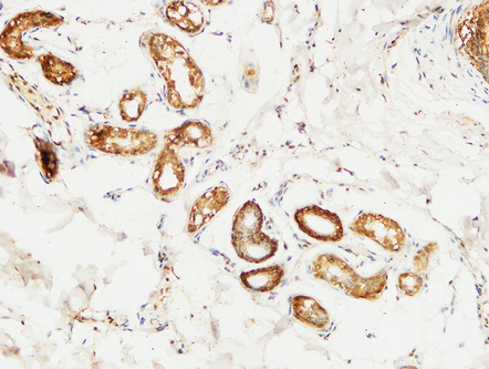
- Immunohistochemical analysis of paraffin-embedded Human skin. 1, Antibody was diluted at 1:200(4° overnight). 2, High-pressure and temperature EDTA, pH8.0 was used for antigen retrieval. 3,Secondary antibody was diluted at 1:200(room temperature, 30min).
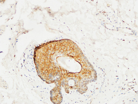
- Immunohistochemical analysis of paraffin-embedded Human skin. 1, Antibody was diluted at 1:200(4° overnight). 2, High-pressure and temperature EDTA, pH8.0 was used for antigen retrieval. 3,Secondary antibody was diluted at 1:200(room temperature, 30min).
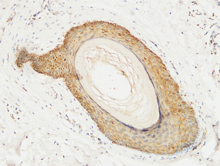
- Immunohistochemical analysis of paraffin-embedded Human skin. 1, Antibody was diluted at 1:200(4° overnight). 2, High-pressure and temperature EDTA, pH8.0 was used for antigen retrieval. 3,Secondary antibody was diluted at 1:200(room temperature, 30min).
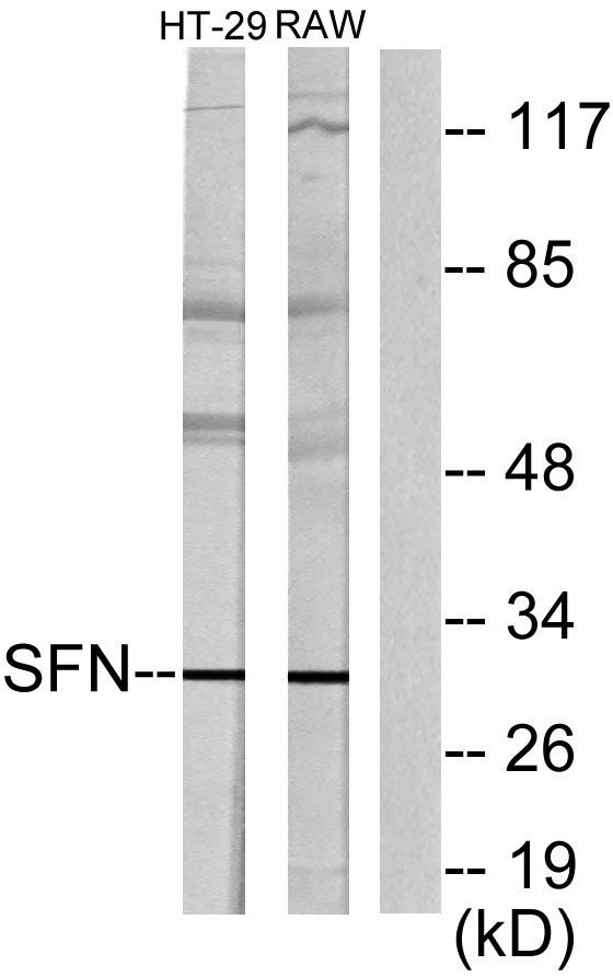
- Western blot analysis of lysates from HT29 cells and RAW264.7 cells, using 14-3-3 sigma Antibody. The lane on the right is blocked with the synthesized peptide.


