Mcl-1 Monoclonal Antibody
- Catalog No.:YM0430
- Applications:WB;IHC;IF;ELISA
- Reactivity:Human
- Target:
- Mcl-1
- Fields:
- >>PI3K-Akt signaling pathway;>>Apoptosis;>>JAK-STAT signaling pathway;>>MicroRNAs in cancer
- Gene Name:
- MCL1
- Protein Name:
- Induced myeloid leukemia cell differentiation protein Mcl-1
- Human Gene Id:
- 4170
- Human Swiss Prot No:
- Q07820
- Mouse Swiss Prot No:
- P97287
- Immunogen:
- Purified recombinant fragment of human MCL-1 expressed in E. Coli.
- Specificity:
- Mcl-1 Monoclonal Antibody detects endogenous levels of Mcl-1 protein.
- Formulation:
- Liquid in PBS containing 50% glycerol, 0.5% BSA and 0.02% sodium azide.
- Source:
- Monoclonal, Mouse
- Dilution:
- WB 1:500 - 1:2000. IHC 1:200 - 1:1000. IF 1:200 - 1:1000. ELISA: 1:10000. Not yet tested in other applications.
- Purification:
- Affinity purification
- Concentration:
- 1 mg/ml
- Storage Stability:
- -15°C to -25°C/1 year(Do not lower than -25°C)
- Other Name:
- MCL1;BCL2L3;Induced myeloid leukemia cell differentiation protein Mcl-1;Bcl-2-like protein 3;Bcl2-L-3;Bcl-2-related protein EAT/mcl1;mcl1/EAT
- Observed Band(KD):
- About 40kd in human,39kd in mouse and rat
- References:
- 1. Ota, N. et al. J. Hum. Genet. 2000. 46: 254-269.
2. Schwertfeger KL, Ryder JW, Anderson SM J Mammary Gland Biol Neoplasia 2000, 3 : 236-251.
- Background:
- This gene encodes an anti-apoptotic protein, which is a member of the Bcl-2 family. Alternative splicing results in multiple transcript variants. The longest gene product (isoform 1) enhances cell survival by inhibiting apoptosis while the alternatively spliced shorter gene products (isoform 2 and isoform 3) promote apoptosis and are death-inducing. [provided by RefSeq, Oct 2010],
- Function:
- function:Involved in the regulation of apoptosis versus cell survival, and in the maintenance of viability but not of proliferation. Mediates its effects by interactions with a number of other regulators of apoptosis. Isoform 1 inhibits apoptosis while isoform 2 promotes it.,induction:Expression increases early during phorbol-ester induced differentiation along the monocyte/macrophage pathway in myeloid leukemia cell lines ML-1. Rapidly up-regulated by CSF2 in ML-1 cells. Up-regulated by heat-shock induced differentiation. Expression increases early during retinoic acid-induced differentiation.,PTM:Cleaved by CASP3 during apoptosis. In intact cells cleavage occurs preferentially after Asp-127, yielding a pro-apoptotic 28 kDa C-terminal fragment.,PTM:Phosphorylated on Thr-163. Treatment with taxol or okadaic acid induces phosphorylation on additional sites.,PTM:Rapidly degraded in the abs
- Subcellular Location:
- Membrane ; Single-pass membrane protein . Cytoplasm. Mitochondrion. Nucleus, nucleoplasm. Cytoplasmic, associated with mitochondria.
- Expression:
- Ewing sarcoma,Mammary gland,Myeloid leukemia cell,Neuroblastoma,Placenta,Th
- June 19-2018
- WESTERN IMMUNOBLOTTING PROTOCOL
- June 19-2018
- IMMUNOHISTOCHEMISTRY-PARAFFIN PROTOCOL
- June 19-2018
- IMMUNOFLUORESCENCE PROTOCOL
- September 08-2020
- FLOW-CYTOMEYRT-PROTOCOL
- May 20-2022
- Cell-Based ELISA│解您多样本WB检测之困扰
- July 13-2018
- CELL-BASED-ELISA-PROTOCOL-FOR-ACETYL-PROTEIN
- July 13-2018
- CELL-BASED-ELISA-PROTOCOL-FOR-PHOSPHO-PROTEIN
- July 13-2018
- Antibody-FAQs
- Products Images
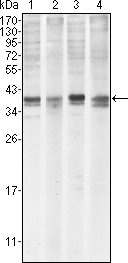
- Western Blot analysis using Mcl-1 Monoclonal Antibody against HeLa (1), BCBL-1 (2), Jurkat (3) and HL60 (4) cell lysate.
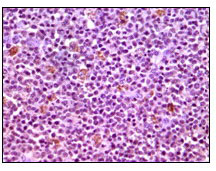
- Immunohistochemistry analysis of paraffin-embedded human lymphnode tissues with DAB staining using Mcl-1 Monoclonal Antibody.

- Confocal immunofluorescence analysis of HepG2 cells using Mcl-1 Monoclonal Antibody (green). Red: Actin filaments have been labeled with DY-554 phalloidin. Blue: DRAQ5 fluorescent DNA dye.


