MPP9 Polyclonal Antibody
- Catalog No.:YT2824
- Applications:WB;IHC
- Reactivity:Human;Rat;Mouse;
- Target:
- MPP9
- Gene Name:
- MPHOSPH9
- Protein Name:
- M-phase phosphoprotein 9
- Human Gene Id:
- 10198
- Human Swiss Prot No:
- Q99550
- Mouse Swiss Prot No:
- A6H5Y1
- Immunogen:
- The antiserum was produced against synthesized peptide derived from human MPHOSPH9. AA range:539-588
- Specificity:
- MPP9 Polyclonal Antibody detects endogenous levels of MPP9 protein.
- Formulation:
- Liquid in PBS containing 50% glycerol, 0.5% BSA and 0.02% sodium azide.
- Source:
- Polyclonal, Rabbit,IgG
- Dilution:
- WB 1:500-2000;IHC 1:50-300
- Purification:
- The antibody was affinity-purified from rabbit antiserum by affinity-chromatography using epitope-specific immunogen.
- Concentration:
- 1 mg/ml
- Storage Stability:
- -15°C to -25°C/1 year(Do not lower than -25°C)
- Other Name:
- MPHOSPH9;MPP9;M-phase phosphoprotein 9
- Observed Band(KD):
- 78kD
- Background:
- PTM:Phosphorylated in M (mitotic) phase.,
- Function:
- PTM:Phosphorylated in M (mitotic) phase.,
- Subcellular Location:
- Cytoplasm, cytoskeleton, microtubule organizing center, centrosome, centriole . Golgi apparatus membrane ; Peripheral membrane protein . Cytoplasm, cytoskeleton, microtubule organizing center, centrosome . Localizes to the distal and proximal end of centriole pairs in duplicated centrosomes. In ciliated cells, localizes to the distal and proximal end of daughter centriole and proximal of the mother centriole but not in the distal end of the mother centriole (PubMed:21399614). Recruited by KIF24 to the distal end of mother centriole where it forms a ring-like structure (PubMed:30375385). .
- Expression:
- Lymphoblast,Tongue,
- June 19-2018
- WESTERN IMMUNOBLOTTING PROTOCOL
- June 19-2018
- IMMUNOHISTOCHEMISTRY-PARAFFIN PROTOCOL
- June 19-2018
- IMMUNOFLUORESCENCE PROTOCOL
- September 08-2020
- FLOW-CYTOMEYRT-PROTOCOL
- May 20-2022
- Cell-Based ELISA│解您多样本WB检测之困扰
- July 13-2018
- CELL-BASED-ELISA-PROTOCOL-FOR-ACETYL-PROTEIN
- July 13-2018
- CELL-BASED-ELISA-PROTOCOL-FOR-PHOSPHO-PROTEIN
- July 13-2018
- Antibody-FAQs
- Products Images
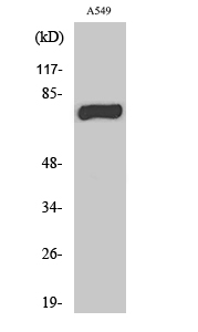
- Western Blot analysis of various cells using MPP9 Polyclonal Antibody
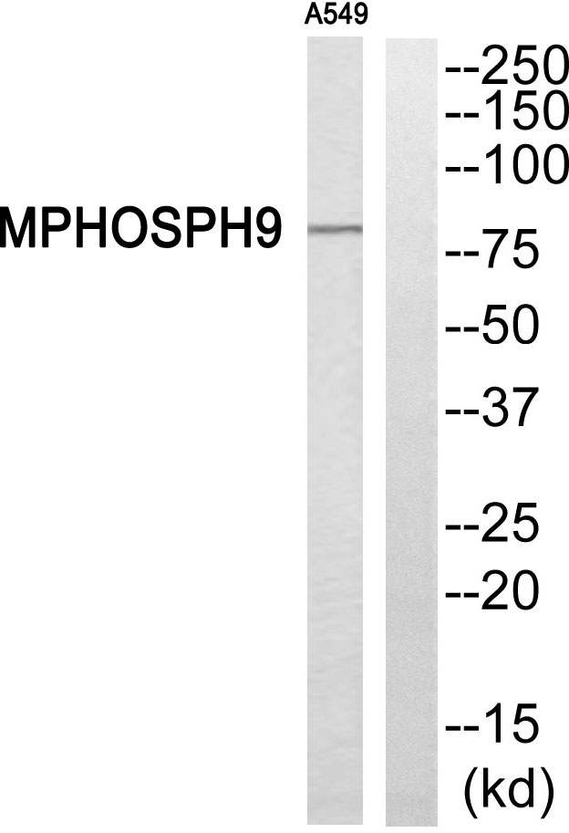
- Western blot analysis of MPHOSPH9 Antibody. The lane on the right is blocked with the MPHOSPH9 peptide.
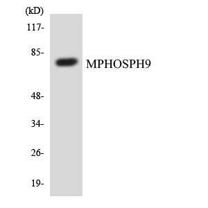
- Western blot analysis of the lysates from COLO205 cells using MPHOSPH9 antibody.
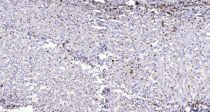
- Immunohistochemical analysis of paraffin-embedded human liver cancer. 1, Antibody was diluted at 1:200(4° overnight). 2, Tris-EDTA,pH9.0 was used for antigen retrieval. 3,Secondary antibody was diluted at 1:200(room temperature, 45min).


