PI 3 kinase p85α (phospho Tyr607) Polyclonal Antibody
- Catalog No.:YP0765
- Applications:IF;WB;IHC;ELISA
- Reactivity:Human;Mouse;Rat;Chicken(testedbyourcustomer)
- Target:
- PI3 Kinase P85α
- Fields:
- >>EGFR tyrosine kinase inhibitor resistance;>>Endocrine resistance;>>Platinum drug resistance;>>ErbB signaling pathway;>>Ras signaling pathway;>>Rap1 signaling pathway;>>cAMP signaling pathway;>>Chemokine signaling pathway;>>HIF-1 signaling pathway;>>FoxO signaling pathway;>>Phosphatidylinositol signaling system;>>Sphingolipid signaling pathway;>>Phospholipase D signaling pathway;>>Autophagy - animal;>>mTOR signaling pathway;>>PI3K-Akt signaling pathway;>>AMPK signaling pathway;>>Apoptosis;>>Longevity regulating pathway;>>Longevity regulating pathway - multiple species;>>Cellular senescence;>>Axon guidance;>>VEGF signaling pathway;>>Osteoclast differentiation;>>Focal adhesion;>>Signaling pathways regulating pluripotency of stem cells;>>Platelet activation;>>Neutrophil extracellular trap formation;>>Toll-like receptor signaling pathway;>>C-type lectin receptor signaling pathway;>>JAK-STAT signaling pathway;>>Natural killer cell mediated cytotoxicity;>>T cell receptor signaling pathway;>
- Gene Name:
- PIK3R1
- Protein Name:
- Phosphatidylinositol 3-kinase regulatory subunit alpha
- Human Gene Id:
- 5295
- Human Swiss Prot No:
- P27986
- Mouse Gene Id:
- 18708
- Mouse Swiss Prot No:
- P26450
- Rat Gene Id:
- 25513
- Rat Swiss Prot No:
- Q63787
- Immunogen:
- The antiserum was produced against synthesized peptide derived from human PI3-kinase p85-alpha around the phosphorylation site of Tyr607. AA range:573-622
- Specificity:
- Phospho-PI 3-kinase p85α (Y607) Polyclonal Antibody detects endogenous levels of PI 3-kinase p85α protein only when phosphorylated at Y607.
- Formulation:
- Liquid in PBS containing 50% glycerol, 0.5% BSA and 0.02% sodium azide.
- Source:
- Polyclonal, Rabbit,IgG
- Dilution:
- IF 1:50-200 WB 1:500 - 1:2000. IHC 1:100 - 1:300. ELISA: 1:10000. Not yet tested in other applications.
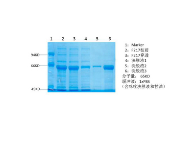
- Purification:
- The antibody was affinity-purified from rabbit antiserum by affinity-chromatography using epitope-specific immunogen.
- Concentration:
- 1 mg/ml
- Storage Stability:
- -15°C to -25°C/1 year(Do not lower than -25°C)
- Other Name:
- PIK3R1;GRB1;Phosphatidylinositol 3-kinase regulatory subunit alpha;PI3-kinase regulatory subunit alpha;PI3K regulatory subunit alpha;PtdIns-3-kinase regulatory subunit alpha;Phosphatidylinositol 3-kinase 85 kDa regulatory subunit alpha;PI3-kinase subunit p85-alpha;PtdIns-3-kinase regulatory subunit p85-alpha
- Observed Band(KD):
- 80kD
- Background:
- Phosphatidylinositol 3-kinase phosphorylates the inositol ring of phosphatidylinositol at the 3-prime position. The enzyme comprises a 110 kD catalytic subunit and a regulatory subunit of either 85, 55, or 50 kD. This gene encodes the 85 kD regulatory subunit. Phosphatidylinositol 3-kinase plays an important role in the metabolic actions of insulin, and a mutation in this gene has been associated with insulin resistance. Alternative splicing of this gene results in four transcript variants encoding different isoforms. [provided by RefSeq, Jun 2011],
- Function:
- disease:Defects in PIK3R1 are a cause of severe insulin resistance.,domain:The SH3 domain mediates the binding to CBLB, and to HIV-1 Nef.,function:Binds to activated (phosphorylated) protein-Tyr kinases, through its SH2 domain, and acts as an adapter, mediating the association of the p110 catalytic unit to the plasma membrane. Necessary for the insulin-stimulated increase in glucose uptake and glycogen synthesis in insulin-sensitive tissues.,PTM:Polyubiquitinated in T-cells by CBLB; which does not promote proteasomal degradation but impairs association with CD28 and CD3Z upon T-cell activation.,similarity:Belongs to the PI3K p85 subunit family.,similarity:Contains 1 Rho-GAP domain.,similarity:Contains 1 SH3 domain.,similarity:Contains 2 SH2 domains.,subunit:Heterodimer of a p110 (catalytic) and a p85 (regulatory) subunits. Interacts with phosphorylated TOM1L1. Interacts with phosphorylat
- Subcellular Location:
- nucleus,cytoplasm,cis-Golgi network,cytosol,plasma membrane,cell-cell junction,phosphatidylinositol 3-kinase complex,phosphatidylinositol 3-kinase complex, class IA,membrane,perinuclear endoplasmic reticulum membrane,
- Expression:
- Isoform 2 is expressed in skeletal muscle and brain, and at lower levels in kidney and cardiac muscle. Isoform 2 and isoform 4 are present in skeletal muscle (at protein level).
Combination of chloroquine diphosphate and salidroside induces human liver cell apoptosis via regulation of mitochondrial dysfunction and autophagy Molecular Medicine Reports Bing Jiang, Longfei Feng, Tao Yang, Wenjing Guo, Yangyang Li, Tao Wang, Chengguang Liu, Haixiang Su WB Human 97H cell
Licoflavone A Suppresses Gastric Cancer Growth and Metastasis by Blocking the VEGFR-2 Signaling Pathway. Journal of Oncology2022;2022:5497991. Human 1 : 1000 MKN-45 cell
The role of the LncRNA-FA2H-2-MLKL pathway in atherosclerosis by regulation of autophagy flux and inflammation through mTOR-dependent signaling. CELL DEATH AND DIFFERENTIATION 2019 Jan 25 WB Human ECs ,SMCs
Morphine promotes the malignant biological behavior of non-small cell lung cancer cells through the MOR/Src/mTOR pathway. Cancer Cell International Cancer Cell Int. 2021 Dec;21(1):1-14 WB Human,Mouse H460 cell-Xenograft H460 human non-small cell lung cancer cells
Isorhamnetin Promotes MKN-45 Gastric Cancer Cell Apoptosis by Inhibiting PI3K-Mediated Adaptive Autophagy in a Hypoxic Environment. JOURNAL OF AGRICULTURAL AND FOOD CHEMISTRY J Agr Food Chem. 2021;XXXX(XXX):XXX-XXX WB Human MKN-45 cells
PM2.5 induces autophagy-mediated cell death via NOS2 signaling in human bronchial epithelium cells. International Journal of Biological Sciences Int J Biol Sci. 2018; 14(5): 557–564 WB Human BEASE-2B cell
The Effects of Maternal Atrazine Exposure and Swimming Training on Spatial Learning Memory and Hippocampal Morphology in Offspring Male Rats via PSD95/NR2B Signaling Pathway. CELLULAR AND MOLECULAR NEUROBIOLOGY Cell Mol Neurobiol. 2019 Oct;39(7):1003-1015 WB Rat 1:500 hippocampus
Luteolin induces apoptosis in vitro through suppressing the MAPK and PI3K signaling pathways in gastric cancer. Oncology Letters Oncol Lett. 2017 Aug;14(2):1993-2000 WB Human 1:1000 BGC-823 cell
Lu, Xueying, et al. "Luteolin induces apoptosis in vitro through suppressing the MAPK and PI3K signaling pathways in gastric cancer." Oncology letters 14.2 (2017): 1993-2000.
5‐FU inhibits migration and invasion of CRC cells through PI3K/AKT pathway regulated by MARCH1. CELL BIOLOGY INTERNATIONAL Cell Biol Int. 2021 Feb;45(2):368-381 WB Human Colorectal cancer(CRC ) tissues sw480s,DLD-1s,NCM460s
Crosstalk between autophagy inhibitor and salidroside-induced apoptosis: A novel strategy for autophagy-based treatment of hepatocellular cancer. INTERNATIONAL IMMUNOPHARMACOLOGY Haixiang Su WB Human 1:500 HepG2 cell,97H cell
Danhong formula alleviates endothelial dysfunction and reduces blood pressure in hypertension by regulating MicroRNA 24 - Phosphatidylinositol 3-Kinase-Serine/Threonine Kinase- Endothelial Nitric Oxide Synthase axis JOURNAL OF ETHNOPHARMACOLOGY Xiaohu Yang WB,IHC Rat 1:50,1:200 aortic tissue
The gut microbiota contributes to the infection of bovine viral diarrhea virus in mice JOURNAL OF VIROLOGY Zecai Zhang WB Mouse peripheral blood lymphocytes (PBL)
- June 19-2018
- WESTERN IMMUNOBLOTTING PROTOCOL
- June 19-2018
- IMMUNOHISTOCHEMISTRY-PARAFFIN PROTOCOL
- June 19-2018
- IMMUNOFLUORESCENCE PROTOCOL
- September 08-2020
- FLOW-CYTOMEYRT-PROTOCOL
- May 20-2022
- Cell-Based ELISA│解您多样本WB检测之困扰
- July 13-2018
- CELL-BASED-ELISA-PROTOCOL-FOR-ACETYL-PROTEIN
- July 13-2018
- CELL-BASED-ELISA-PROTOCOL-FOR-PHOSPHO-PROTEIN
- July 13-2018
- Antibody-FAQs
- Products Images
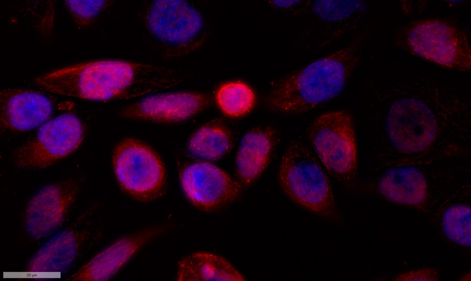
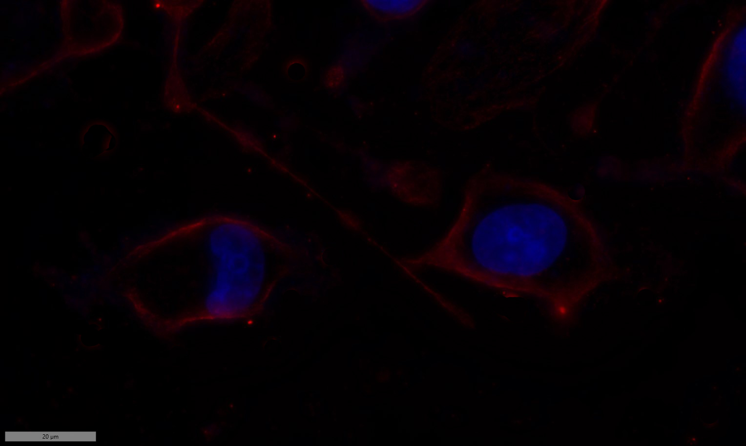
- Immunofluorescence analysis of Siha cell. 1,primary Antibody was diluted at 1:100(4°C overnight). 2, Goat Anti Rabbit IgG (H&L) - AFluor 594 Secondary antibody(catalog No: RS3611) was diluted at 1:500(room temperature, 50min).

- Lu, Xueying, et al. "Luteolin induces apoptosis in vitro through suppressing the MAPK and PI3K signaling pathways in gastric cancer." Oncology letters 14.2 (2017): 1993-2000.
-if-137.jpg)
- Immunofluorescence analysis of rat-heart tissue. 1,PI 3-kinase p85α (phospho Tyr607) Polyclonal Antibody(red) was diluted at 1:200(4°C,overnight). 2, Cy3 labled Secondary antibody was diluted at 1:300(room temperature, 50min).3, Picture B: DAPI(blue) 10min. Picture A:Target. Picture B: DAPI. Picture C: merge of A+B
-if-138.jpg)
- Immunofluorescence analysis of rat-heart tissue. 1,PI 3-kinase p85α (phospho Tyr607) Polyclonal Antibody(red) was diluted at 1:200(4°C,overnight). 2, Cy3 labled Secondary antibody was diluted at 1:300(room temperature, 50min).3, Picture B: DAPI(blue) 10min. Picture A:Target. Picture B: DAPI. Picture C: merge of A+B
poly-ihc-human-liver.jpg)
- Immunohistochemical analysis of paraffin-embedded Human-liver tissue. 1,PI 3-kinase p85α (phospho Tyr607) Polyclonal Antibody was diluted at 1:200(4°C,overnight). 2, Sodium citrate pH 6.0 was used for antibody retrieval(>98°C,20min). 3,Secondary antibody was diluted at 1:200(room tempeRature, 30min). Negative control was used by secondary antibody only.
poly-ihc-human-liver-cancer.jpg)
- Immunohistochemical analysis of paraffin-embedded Human-liver-cancer tissue. 1,PI 3-kinase p85α (phospho Tyr607) Polyclonal Antibody was diluted at 1:200(4°C,overnight). 2, Sodium citrate pH 6.0 was used for antibody retrieval(>98°C,20min). 3,Secondary antibody was diluted at 1:200(room tempeRature, 30min). Negative control was used by secondary antibody only.
poly-ihc-human-stomach-cancer.jpg)
- Immunohistochemical analysis of paraffin-embedded Human-stomach-cancer tissue. 1,PI 3-kinase p85α (phospho Tyr607) Polyclonal Antibody was diluted at 1:200(4°C,overnight). 2, Sodium citrate pH 6.0 was used for antibody retrieval(>98°C,20min). 3,Secondary antibody was diluted at 1:200(room tempeRature, 30min). Negative control was used by secondary antibody only.
poly-ihc-rat-heart.jpg)
- Immunohistochemical analysis of paraffin-embedded Rat-heart tissue. 1,PI 3-kinase p85α (phospho Tyr607) Polyclonal Antibody was diluted at 1:200(4°C,overnight). 2, Sodium citrate pH 6.0 was used for antibody retrieval(>98°C,20min). 3,Secondary antibody was diluted at 1:200(room tempeRature, 30min). Negative control was used by secondary antibody only.
poly-ihc-rat-spinal-cord.jpg)
- Immunohistochemical analysis of paraffin-embedded Rat-spinal-cord tissue. 1,PI 3-kinase p85α (phospho Tyr607) Polyclonal Antibody was diluted at 1:200(4°C,overnight). 2, Sodium citrate pH 6.0 was used for antibody retrieval(>98°C,20min). 3,Secondary antibody was diluted at 1:200(room tempeRature, 30min). Negative control was used by secondary antibody only.
poly-ihc-rat-brain.jpg)
- Immunohistochemical analysis of paraffin-embedded Rat-brain tissue. 1,PI 3-kinase p85α (phospho Tyr607) Polyclonal Antibody was diluted at 1:200(4°C,overnight). 2, Sodium citrate pH 6.0 was used for antibody retrieval(>98°C,20min). 3,Secondary antibody was diluted at 1:200(room tempeRature, 30min). Negative control was used by secondary antibody only.
poly-ihc-mouse-colon.jpg)
- Immunohistochemical analysis of paraffin-embedded Mouse-colon tissue. 1,PI 3-kinase p85α (phospho Tyr607) Polyclonal Antibody was diluted at 1:200(4°C,overnight). 2, Sodium citrate pH 6.0 was used for antibody retrieval(>98°C,20min). 3,Secondary antibody was diluted at 1:200(room tempeRature, 30min). Negative control was used by secondary antibody only.
poly-ihc-mouse-brain.jpg)
- Immunohistochemical analysis of paraffin-embedded Mouse-brain tissue. 1,PI 3-kinase p85α (phospho Tyr607) Polyclonal Antibody was diluted at 1:200(4°C,overnight). 2, Sodium citrate pH 6.0 was used for antibody retrieval(>98°C,20min). 3,Secondary antibody was diluted at 1:200(room tempeRature, 30min). Negative control was used by secondary antibody only.
poly-ihc-mouse-spleen.jpg)
- Immunohistochemical analysis of paraffin-embedded Mouse-spleen tissue. 1,PI 3-kinase p85α (phospho Tyr607) Polyclonal Antibody was diluted at 1:200(4°C,overnight). 2, Sodium citrate pH 6.0 was used for antibody retrieval(>98°C,20min). 3,Secondary antibody was diluted at 1:200(room tempeRature, 30min). Negative control was used by secondary antibody only.
- Western Blot analysis of various cells using Phospho-PI 3-kinase p85α (Y607) Polyclonal Antibody diluted at 1:1000
.jpg)
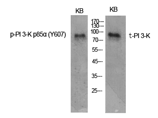
- Western Blot analysis of KB cells using Phospho-PI 3-kinase p85α (Y607) Polyclonal Antibody diluted at 1:1000
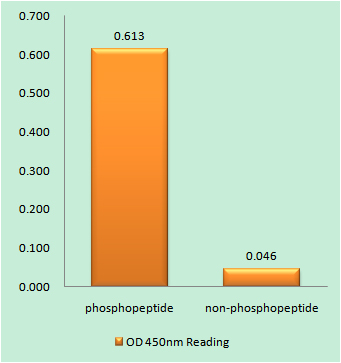
- Enzyme-Linked Immunosorbent Assay (Phospho-ELISA) for Immunogen Phosphopeptide (Phospho-left) and Non-Phosphopeptide (Phospho-right), using PI3-kinase p85-alpha (Phospho-Tyr607) Antibody

- Immunohistochemistry analysis of paraffin-embedded human brain, using PI3-kinase p85-alpha (Phospho-Tyr607) Antibody. The picture on the right is blocked with the phospho peptide.
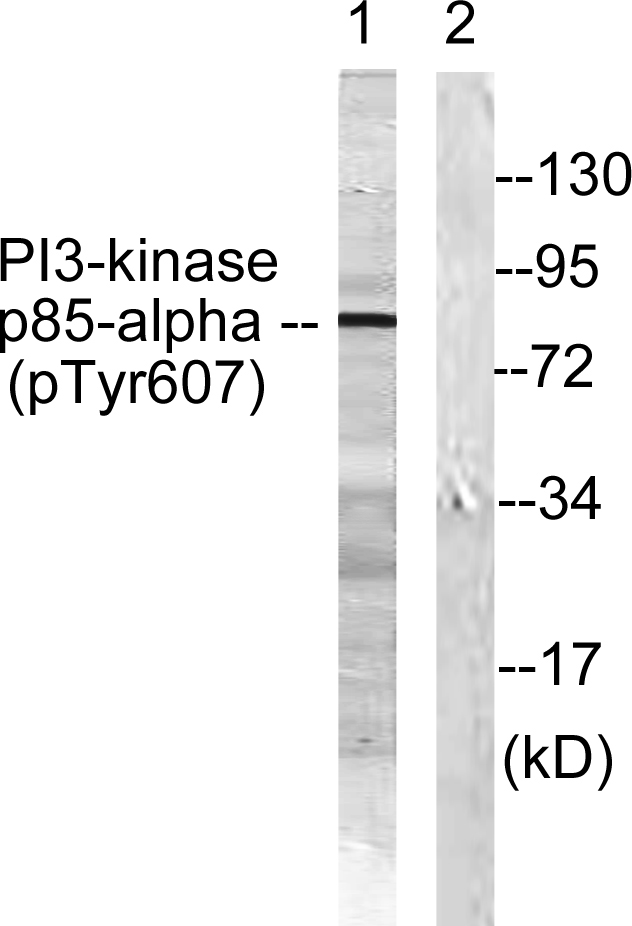
- Western blot analysis of lysates from rat kidney, using PI3-kinase p85-alpha (Phospho-Tyr607) Antibody. The lane on the right is blocked with the phospho peptide.

.jpg)