- Home
- About
- Promotions
-
Products
-
Elisa Kits
- |
-
Primary antibodies
- |
-
Secondary antibodies
- |
-
Proteins
- |
-
IHC reagents
- |
-
WB reagents
- PonceauS Staining Solution
- PBST Washing Buffer, 10X
- 1.5M Tris-HCl Buffer, pH8.8
- 1M Tris-HCl Buffer, pH6.8
- 10% SDS Solution
- Prestained Protein Marker
- TBST Washing Buffer, 10X
- SDS PAGE Loading Buffer, 5X
- Stripping Buffered Solution
- Tris Buffer, pH7.4, 10X
- Total Protein Extraction Kit
- Running Buffer, 10X
- Transfer Buffer, 10X
- 30% Acr-Bis(29:1) Solution
- Tris电泳液速溶颗粒
- PBS(1X, premixed powder)
- TBS(1X, premixed powder)
- 快速封闭液
- 转膜液速溶颗粒
- Chemical reagents
- News
- Distributor
- Resources
- Contact
- Home
- >
- Info
- >
- 鼠兔六标七色荧光检测试剂盒
- >
- Go Back
鼠兔六标七色荧光检测试剂盒
- Catalog No.:RS0039
- Applications:IF/mIHC
- Storage Stability:
- See datasheet
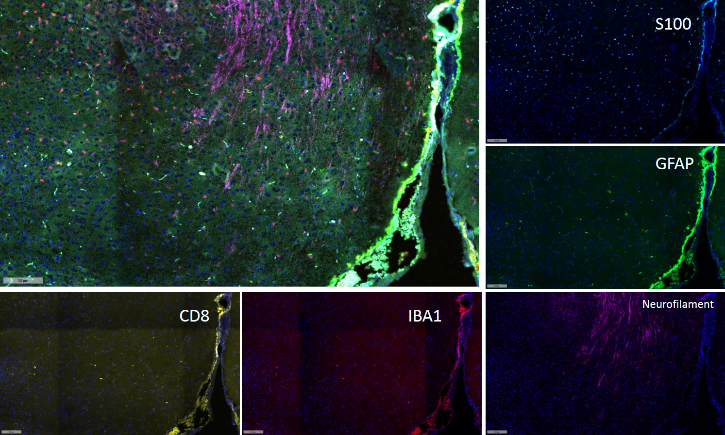
- Fluorescence multiplex immunohistochemical analysis of Mouse brain tissue (formalin-fixed paraffin-embedded section).
The immunostaining was performed by Sextuple-Fluorescence kit (RS0039, Immunoway). GFAP mouse mAb(YM4426 Immunoway) green, S100 mouse mAb(YM6987 Immunoway) cyan,Neurofilament mouse mAb(YM6897 Immunoway) purple,Iba 1 mouse mAb(YM4765 Immunoway) red,CD8 a mouse mAb(YM4815 Immunoway) yellow,
The section was incubated in 5 rounds of staining; sequentially for Anti-antibodies; each using a separate fluorescent tyramide signal amplification system. EDTA based antigen retrieval (Immunoway YS0004, pH 9.0, 20 minutes) was used in between rounds of tyramide signal amplification to remove the antibody from the previous round, to avoid any cross-reactivity. DAPI (dark blue) was used as a nuclear counter stain.
Microscopy and pseudocoloring of individual dyes was performed using a Slideviewer Imaging System (Excilone).

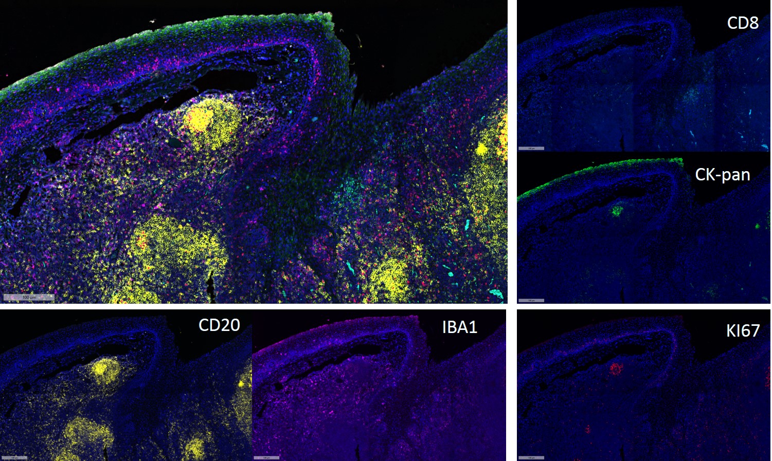
- Fluorescence multiplex immunohistochemical analysis of Mouse brain tissue (formalin-fixed paraffin-embedded section). The immunostaining was performed by Sextuple-Fluorescence kit (RS0039, Immunoway).Human colon carcinoma was tested by S100,CD4, CD8,CD68,PDL1,CKpan mouse mAb with 6 different TSA Fluorescence regent. Numbers of functional cells(S100,CD4, CD8,CD68,PDL1) was calculated at every 5μm within 100μm range around each tumor cell(CKpan+).
Microscopy and pseudocoloring of individual dyes was performed using a Slideviewer Imaging System (Excilone).