


Catalog: YT0079
Size
Price
Status
Qty.
200μL
$450.00
In stock
0
100μL
$280.00
In stock
0
40μL
$150.00
In stock
0
Add to cart


Collected
Collect
Main Information
Target
AChE
Host Species
Rabbit
Reactivity
Human, Mouse, Rat
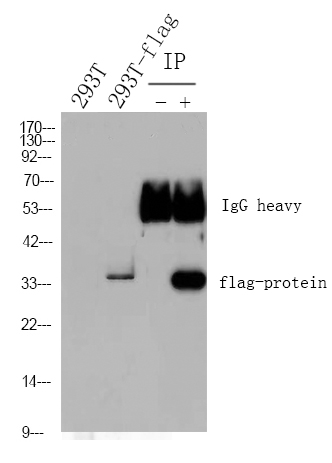
Applications
WB, ELISA
MW
70kD (Observed)
Conjugate/Modification
Unmodified
Detailed Information
Recommended Dilution Ratio
WB 1:500-1:2000; ELISA 1:5000; Not yet tested in other applications.
Formulation
Liquid in PBS containing 50% glycerol, 0.5% BSA and 0.02% sodium azide.
Specificity
AChE Polyclonal Antibody detects endogenous levels of AChE protein.
Purification
The antibody was affinity-purified from rabbit antiserum by affinity-chromatography using epitope-specific immunogen.
Storage
-15°C to -25°C/1 year(Do not lower than -25°C)
Concentration
1 mg/ml
MW(Observed)
70kD
Modification
Unmodified
Clonality
Polyclonal
Isotype
IgG
Related Products
Secondary Antibodies
Goat Anti Mouse IgG(H+L) (HRP)
RS0001
More→
Secondary Antibodies

Goat Anti Rabbit IgG(H+L) (HRP)
RS0002
More→
Primary Antibodies
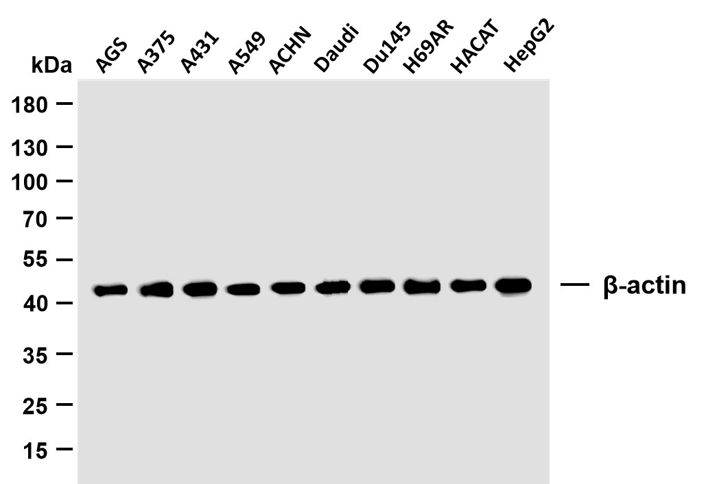
β-actin (PTR2364) Mouse mAb
YM3028
More→
Primary Antibodies
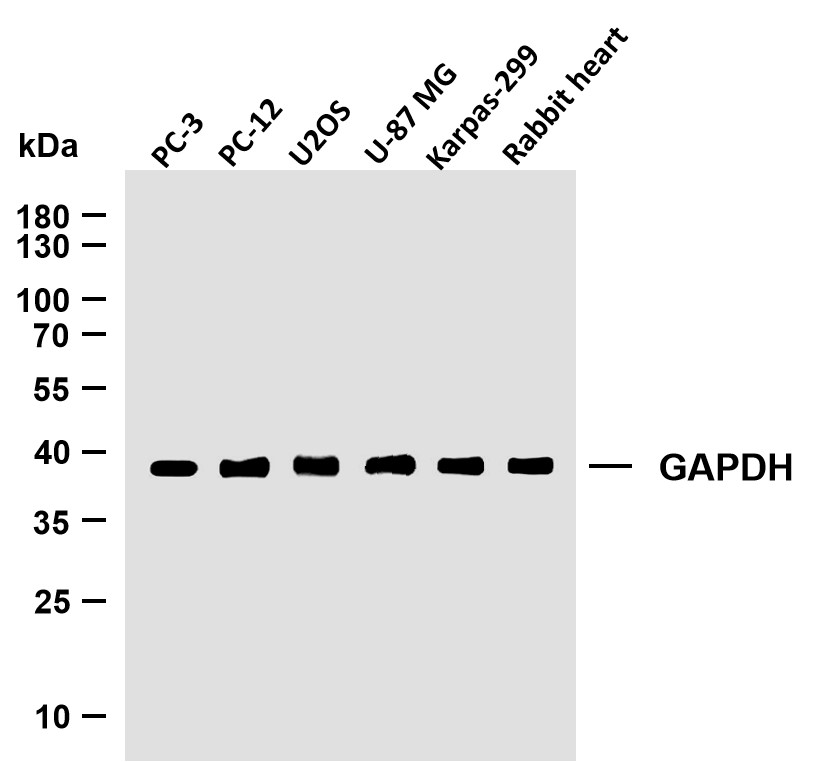
GAPDH (PTR2304) Mouse mAb
YM3029
More→
Antigen&Target Information
Immunogen:
The antiserum was produced against synthesized peptide derived from human ACHE. AA range:551-600
show all
Specificity:
AChE Polyclonal Antibody detects endogenous levels of AChE protein.
show all
Gene Name:
ACHE
show all
Protein Name:
Acetylcholinesterase
show all
Other Name:
ACHE ;
Acetylcholinesterase ;
AChE
Acetylcholinesterase ;
AChE
show all
Background:
Acetylcholinesterase hydrolyzes the neurotransmitter, acetylcholine at neuromuscular junctions and brain cholinergic synapses, and thus terminates signal transmission. It is also found on the red blood cell membranes, where it constitutes the Yt blood group antigen. Acetylcholinesterase exists in multiple molecular forms which possess similar catalytic properties, but differ in their oligomeric assembly and mode of cell attachment to the cell surface. It is encoded by the single ACHE gene, and the structural diversity in the gene products arises from alternative mRNA splicing, and post-translational associations of catalytic and structural subunits. The major form of acetylcholinesterase found in brain, muscle and other tissues is the hydrophilic species, which forms disulfide-linked oligomers with collagenous, or lipid-containing structural subunits. The other, alternatively
show all
Function:
Catalytic activity:Acetylcholine + H(2)O = choline + acetate.,Disease:Behaves as an amyloid-promoting factor to promote the formation of amyloid plaques in Alzheimer disease.,Function:Terminates signal transduction at the neuromuscular junction by rapid hydrolysis of the acetylcholine released into the synaptic cleft. Role in neuronal apoptosis.,online information:Acetylcholinesterase entry,online information:Blood group antigen gene mutation database,polymorphism:ACHE is responsible for the Yt blood group system. The molecular basis of the Yt(a)=Yt1/Yt(b)=Yt2 blood group antigens is a single variation in position 353; His-353 corresponds to Yt(a) and the rare variant with Asn-353 to Yt(b).,similarity:Belongs to the type-B carboxylesterase/lipase family.,subcellular location:Only observed in apoptotic nuclei.,subunit:Interacts with PRIMA1. The interaction with PRIMA1 is required to anchor it to the basal lamina of cells and organize into tetramers (By similarity). Isoform H generates GPI-anchored dimers; disulfide linked. Isoform T generates multiple structures, ranging from monomers and dimers to collagen-tailed and hydrophobic-tailed forms, in which catalytic tetramers are associated with anchoring proteins that attach them to the basal lamina or to cell membranes. In the collagen-tailed forms, isoform T subunits are associated with a specific collagen, COLQ, which triggers the formation of isoform T tetramers, from monomers and dimers. Isoform R may be monomeric.,tissue specificity:Isoform H is highly expressed in erythrocytes.,
show all
Cellular Localization:
Cell junction, synapse . Secreted . Cell membrane ; Peripheral membrane protein .; [Isoform T]: Nucleus. Only observed in apoptotic nuclei.; [Isoform H]: Cell membrane ; Lipid-anchor, GPI-anchor ; Extracellular side .
show all
Tissue Expression:
Isoform H is highly expressed in erythrocytes.
show all
Research Areas:
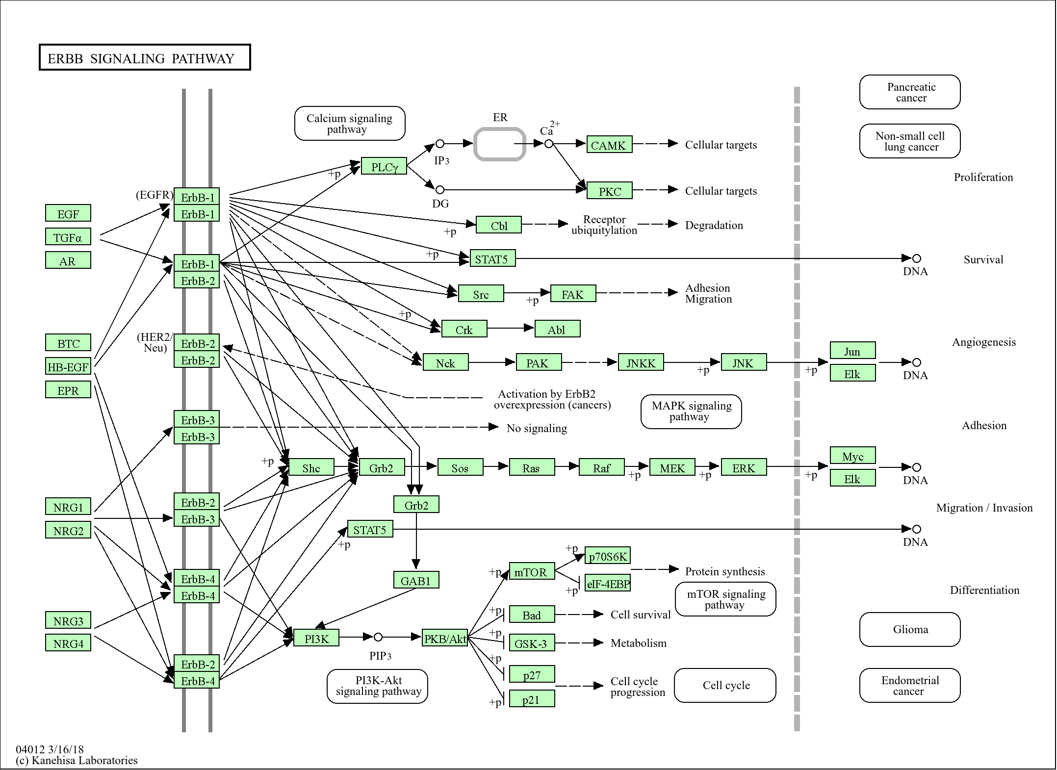
>>Glycerophospholipid metabolism ;
>>Cholinergic synapse
>>Cholinergic synapse
show all
Signaling Pathway
Reference Citation({{totalcount}})
Catalog: YT0079
Size
Price
Status
Qty.
200μL
$450.00
In stock
0
100μL
$280.00
In stock
0
40μL
$150.00
In stock
0
Add to cart
Collected
Collect
Recently Viewed Products
Clear allPRODUCTS
CUSTOMIZED
ABOUT US


Toggle night Mode
{{pinfoXq.title || ''}}
Catalog: {{pinfoXq.catalog || ''}}
Filter:
All
{{item.name}}
{{pinfo.title}}
-{{pinfo.catalog}}
Main Information
Target
{{pinfo.target}}
Reactivity
{{pinfo.react}}
Applications
{{pinfo.applicat}}
Conjugate/Modification
{{pinfo.coupling}}/{{pinfo.modific}}
MW (kDa)
{{pinfo.mwcalc}}
Host Species
{{pinfo.hostspec}}
Isotype
{{pinfo.isotype}}
Product {{index}}/{{pcount}}
Prev
Next
{{pvTitle}}
Scroll wheel zooms the picture
{{pvDescr}}