TGFβ RII Polyclonal Antibody
- Catalog No.:YT4629
- Applications:IF;WB;IHC;ELISA
- Reactivity:Human;Mouse;Rat
- Target:
- TGF β Receptor II
- Fields:
- >>MAPK signaling pathway;>>Cytokine-cytokine receptor interaction;>>FoxO signaling pathway;>>Endocytosis;>>Cellular senescence;>>TGF-beta signaling pathway;>>Osteoclast differentiation;>>Hippo signaling pathway;>>Adherens junction;>>Th17 cell differentiation;>>Relaxin signaling pathway;>>AGE-RAGE signaling pathway in diabetic complications;>>Chagas disease;>>Hepatitis B;>>Human T-cell leukemia virus 1 infection;>>Pathways in cancer;>>Transcriptional misregulation in cancer;>>Colorectal cancer;>>Pancreatic cancer;>>Chronic myeloid leukemia;>>Hepatocellular carcinoma;>>Gastric cancer;>>Diabetic cardiomyopathy
- Gene Name:
- TGFBR2
- Protein Name:
- TGF-beta receptor type-2
- Human Gene Id:
- 7048
- Human Swiss Prot No:
- P37173
- Mouse Gene Id:
- 21813
- Mouse Swiss Prot No:
- Q62312
- Rat Gene Id:
- 81810
- Rat Swiss Prot No:
- P38438
- Immunogen:
- The antiserum was produced against synthesized peptide derived from human TGF beta Receptor II. AA range:91-140
- Specificity:
- TGFβ RII Polyclonal Antibody detects endogenous levels of TGFβ RII protein.
- Formulation:
- Liquid in PBS containing 50% glycerol, 0.5% BSA and 0.02% sodium azide.
- Source:
- Polyclonal, Rabbit,IgG
- Dilution:
- IF 1:50-200 WB 1:500 - 1:2000. IHC 1:100 - 1:300. ELISA: 1:20000. Not yet tested in other applications.
- Purification:
- The antibody was affinity-purified from rabbit antiserum by affinity-chromatography using epitope-specific immunogen.
- Concentration:
- 1 mg/ml
- Storage Stability:
- -15°C to -25°C/1 year(Do not lower than -25°C)
- Other Name:
- TGFBR2;TGF-beta receptor type-2;TGFR-2;TGF-beta type II receptor;Transforming growth factor-beta receptor type II;TGF-beta receptor type II;TbetaR-II
- Observed Band(KD):
- 65kD
- Background:
- This gene encodes a member of the Ser/Thr protein kinase family and the TGFB receptor subfamily. The encoded protein is a transmembrane protein that has a protein kinase domain, forms a heterodimeric complex with another receptor protein, and binds TGF-beta. This receptor/ligand complex phosphorylates proteins, which then enter the nucleus and regulate the transcription of a subset of genes related to cell proliferation. Mutations in this gene have been associated with Marfan Syndrome, Loeys-Deitz Aortic Aneurysm Syndrome, and the development of various types of tumors. Alternatively spliced transcript variants encoding different isoforms have been characterized. [provided by RefSeq, Jul 2008],
- Function:
- catalytic activity:ATP + [receptor-protein] = ADP + [receptor-protein] phosphate.,cofactor:Magnesium or manganese.,disease:Defects in TGFBR2 are a cause of esophageal cancer [MIM:133239].,disease:Defects in TGFBR2 are the cause of aortic aneurysm familial thoracic type 3 (AAT3) [MIM:610380]. Aneurysms and dissections of the aorta usually result from degenerative changes in the aortic wall. Thoracic aortic aneurysms and dissections are primarily associated with a characteristic histologic appearance known as 'medial necrosis' or 'Erdheim cystic medial necrosis' in which there is degeneration and fragmentation of elastic fibers, loss of smooth muscle cells, and an accumulation of basophilic ground substance. AAT3 is an autosomal dominant disorder with reduced penetrance and variable expression.,disease:Defects in TGFBR2 are the cause of hereditary non-polyposis colorectal cancer type 6 (HN
- Subcellular Location:
- Cell membrane ; Single-pass type I membrane protein . Membrane raft .; [Isoform 3]: Secreted .
- Expression:
- Cerebellum,Colon,Epithelium,Glial cell,Liver,
Astragalus Polysaccharide Eases G1 Phase-Correlative Bystander Effects through Mediation of TGF-βR/MAPK/ROS Signal Pathway After Carbon Ion Irradiation in BMSCs. AMERICAN JOURNAL OF CHINESE MEDICINE Am J Chinese Med. 2019 May;47(03):595-612 WB Human BMSCs
M2 macrophage-mediated interleukin-4 signalling induces myofibroblast phenotype during the progression of benign prostatic hyperplasia. Cell Death & Disease Cell Death Dis. 2018 Jul;9(7):1-13 WB Human 1:1000 prostate tissue prostate fibroblasts
Regulation of proliferation and cell cycle by protein regulator of cytokinesis 1 in oral squamous cell carcinoma. Cell Death & Disease 2018 May 11 WB Human 1:500 HSC-2 cell,Cal-27 cell
Pan, Ming-Ming, et al. "Inhibition of TGF-β1/Smad signal pathway is involved in the effect of Cordyceps sinensis against renal fibrosis in 5/6 nephrectomy rats." Food and chemical toxicology 58 (2013): 487-494.
Fu, Na, et al. "Role of LncRNA-activated by transforming growth factor beta in the progression of hepatitis C virus-related liver fibrosis." Discovery medicine 22.119 (2016): 29-42.
- June 19-2018
- WESTERN IMMUNOBLOTTING PROTOCOL
- June 19-2018
- IMMUNOHISTOCHEMISTRY-PARAFFIN PROTOCOL
- June 19-2018
- IMMUNOFLUORESCENCE PROTOCOL
- September 08-2020
- FLOW-CYTOMEYRT-PROTOCOL
- May 20-2022
- Cell-Based ELISA│解您多样本WB检测之困扰
- July 13-2018
- CELL-BASED-ELISA-PROTOCOL-FOR-ACETYL-PROTEIN
- July 13-2018
- CELL-BASED-ELISA-PROTOCOL-FOR-PHOSPHO-PROTEIN
- July 13-2018
- Antibody-FAQs
- Products Images
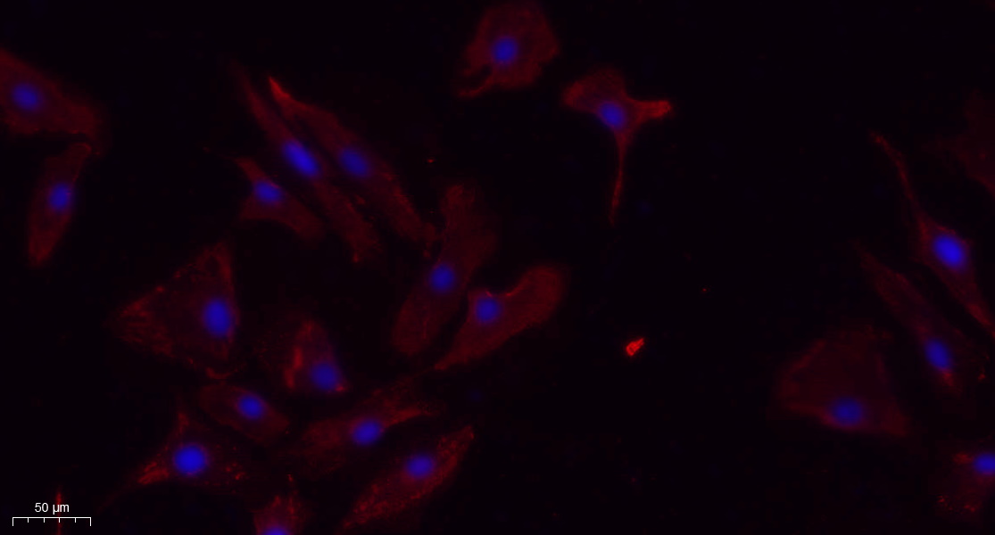
- Immunofluorescence analysis of A549. 1,primary Antibody(red) was diluted at 1:200(4°C overnight). 2, Goat Anti Rabbit IgG (H&L) - Alexa Fluor 594 Secondary antibody was diluted at 1:1000(room temperature, 50min).3, Picture B: DAPI(blue) 10min.
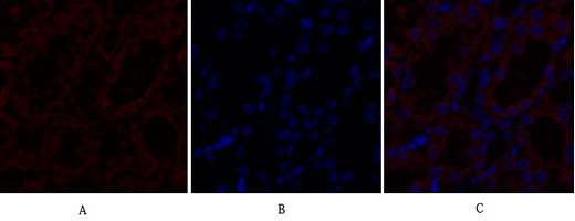
- Immunofluorescence analysis of rat-kidney tissue. 1,TGFβ RII Polyclonal Antibody(red) was diluted at 1:200(4°C,overnight). 2, Cy3 labled Secondary antibody was diluted at 1:300(room temperature, 50min).3, Picture B: DAPI(blue) 10min. Picture A:Target. Picture B: DAPI. Picture C: merge of A+B
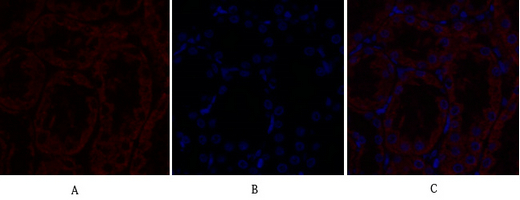
- Immunofluorescence analysis of rat-kidney tissue. 1,TGFβ RII Polyclonal Antibody(red) was diluted at 1:200(4°C,overnight). 2, Cy3 labled Secondary antibody was diluted at 1:300(room temperature, 50min).3, Picture B: DAPI(blue) 10min. Picture A:Target. Picture B: DAPI. Picture C: merge of A+B
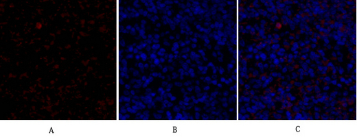
- Immunofluorescence analysis of rat-spleen tissue. 1,TGFβ RII Polyclonal Antibody(red) was diluted at 1:200(4°C,overnight). 2, Cy3 labled Secondary antibody was diluted at 1:300(room temperature, 50min).3, Picture B: DAPI(blue) 10min. Picture A:Target. Picture B: DAPI. Picture C: merge of A+B
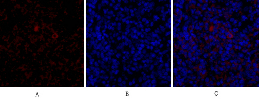
- Immunofluorescence analysis of rat-spleen tissue. 1,TGFβ RII Polyclonal Antibody(red) was diluted at 1:200(4°C,overnight). 2, Cy3 labled Secondary antibody was diluted at 1:300(room temperature, 50min).3, Picture B: DAPI(blue) 10min. Picture A:Target. Picture B: DAPI. Picture C: merge of A+B
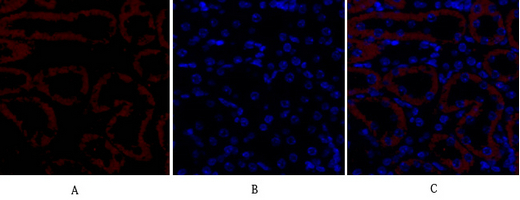
- Immunofluorescence analysis of mouse-kidney tissue. 1,TGFβ RII Polyclonal Antibody(red) was diluted at 1:200(4°C,overnight). 2, Cy3 labled Secondary antibody was diluted at 1:300(room temperature, 50min).3, Picture B: DAPI(blue) 10min. Picture A:Target. Picture B: DAPI. Picture C: merge of A+B

- Immunofluorescence analysis of mouse-kidney tissue. 1,TGFβ RII Polyclonal Antibody(red) was diluted at 1:200(4°C,overnight). 2, Cy3 labled Secondary antibody was diluted at 1:300(room temperature, 50min).3, Picture B: DAPI(blue) 10min. Picture A:Target. Picture B: DAPI. Picture C: merge of A+B
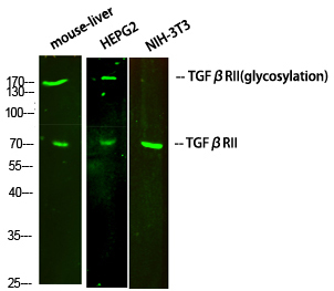
- Western Blot analysis of various cells using primary antibody diluted at 1:1000(4°C overnight). Secondary antibody:Goat Anti-rabbit IgG IRDye 800( diluted at 1:5000, 25°C, 1 hour)

- Western Blot analysis of various cells using TGFβ RII Polyclonal Antibody diluted at 1:2000
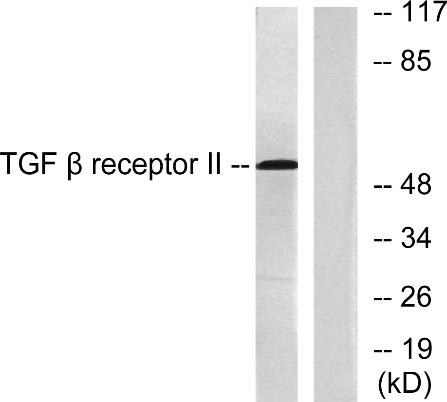
- Western blot analysis of lysates from HepG2 (65K) cells, using TGF beta Receptor II Antibody. The lane on the right is blocked with the synthesized peptide.
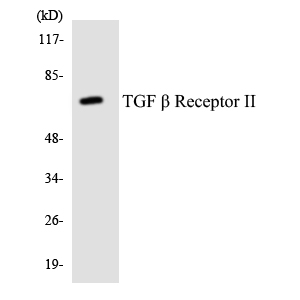
- Western blot analysis of the lysates from HT-29 cells using TGF β Receptor II antibody.
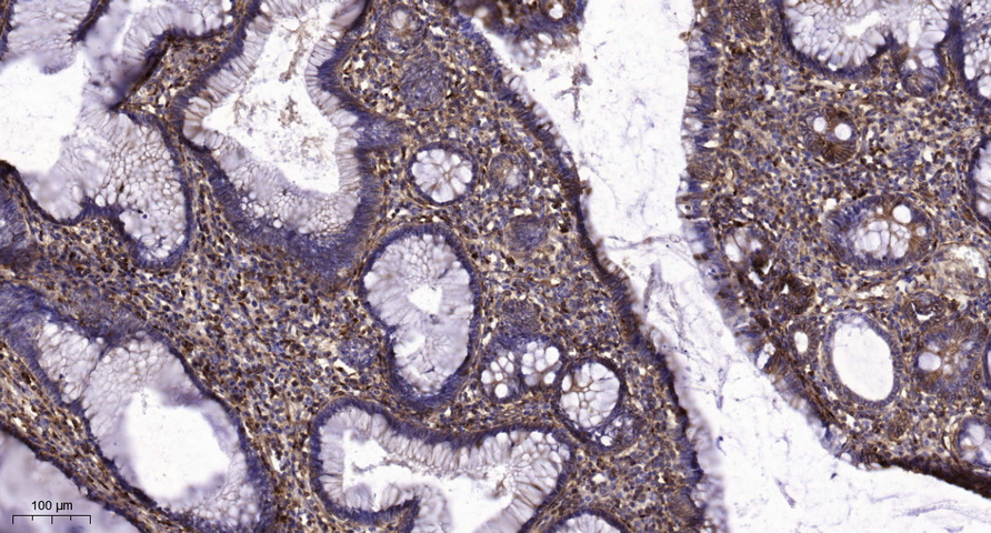
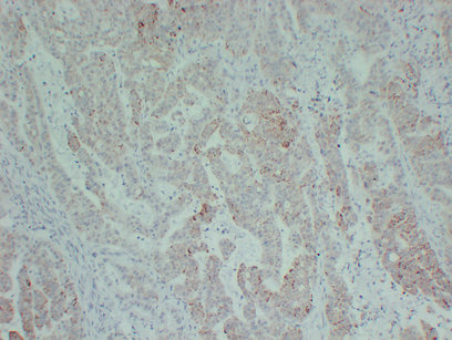
- Immunohistochemical analysis of paraffin-embedded human colon cancer. 1, Tris-EDTA,pH9.0 was used for antigen retrieval. 2 Antibody was diluted at 1:200(4° overnight.3,Secondary antibody was diluted at 1:200(room temperature, 45min).