SNAI 1 Polyclonal Antibody
- Catalog No.:YT4351
- Applications:WB;IP;IHC;IF;ELISA
- Reactivity:Human;Mouse;Monkey
- Target:
- SNAI1
- Fields:
- >>Adherens junction
- Gene Name:
- SNAI1
- Protein Name:
- Zinc finger protein SNAI1(snail)
- Human Gene Id:
- 6615
- Human Swiss Prot No:
- O95863
- Mouse Gene Id:
- 20613
- Mouse Swiss Prot No:
- Q02085
- Immunogen:
- The antiserum was produced against synthesized peptide derived from human SNAI1. AA range:215-264
- Specificity:
- SNAI 1 Polyclonal Antibody detects endogenous levels of SNAI 1 protein.
- Formulation:
- Liquid in PBS containing 50% glycerol, 0.5% BSA and 0.02% sodium azide.
- Source:
- Polyclonal, Rabbit,IgG
- Dilution:
- WB 1:500 - 1:2000. IHC 1:100 - 1:300. IF 1:200 - 1:1000. ELISA: 1:5000. Not yet tested in other applications.
- Purification:
- The antibody was affinity-purified from rabbit antiserum by affinity-chromatography using epitope-specific immunogen.
- Concentration:
- 1 mg/ml
- Storage Stability:
- -15°C to -25°C/1 year(Do not lower than -25°C)
- Other Name:
- SNAI1;SNAH;Zinc finger protein SNAI1;Protein snail homolog 1;Protein sna
- Observed Band(KD):
- 29kD
- Background:
- snail family transcriptional repressor 1(SNAI1) Homo sapiens The Drosophila embryonic protein snail is a zinc finger transcriptional repressor which downregulates the expression of ectodermal genes within the mesoderm. The nuclear protein encoded by this gene is structurally similar to the Drosophila snail protein, and is also thought to be critical for mesoderm formation in the developing embryo. At least two variants of a similar processed pseudogene have been found on chromosome 2. [provided by RefSeq, Jul 2008],
- Function:
- function:Seems to be involved in embryonic mesoderm formation. Binds to 3 E-boxes of the E-cadherin gene promoter and represses its transcription.,similarity:Belongs to the snail C2H2-type zinc-finger protein family.,similarity:Contains 4 C2H2-type zinc fingers.,tissue specificity:Expressed in a variety of tissues with the highest expression in kidney.,
- Subcellular Location:
- Nucleus . Cytoplasm . Once phosphorylated (probably on Ser-107, Ser-111, Ser-115 and Ser-119) it is exported from the nucleus to the cytoplasm where subsequent phosphorylation of the destruction motif and ubiquitination involving BTRC occurs. .
- Expression:
- Expressed in a variety of tissues with the highest expression in kidney. Expressed in mesenchymal and epithelial cell lines.
Afatinib Reverses EMT via Inhibiting CD44-Stat3 Axis to Promote Radiosensitivity in Nasopharyngeal Carcinoma Pharmaceuticals Huichao Huang, Fangling Huang, Xujun Liang, Ying Fu, Zhe Cheng, Yan Huang, Zhuchu Chen, Yankun Duan, Yongheng Chen WB Human 5-8F cell, HNE2 cell
Doublecortin-like kinase 2 promotes breast cancer cell invasion and metastasis Clinical & Translational Oncology He, Yanling, Dai, Xiaoqin, Li, Shengnan, Zhang, Xinyuan, Gong, Kunxiang, Song, Kai, Shi, Jian WB Human MDA-MB-231 cell, MDA-MB-453 cell
Promoter methylation-regulated miR-148a-3p inhibits lung adenocarcinoma (LUAD) progression by targeting MAP3K9. ACTA PHARMACOLOGICA SINICA2022 Apr;:1-10. Human 1:1000 A549 cell, NCI-H1299 cell
Ponicidin inhibits pro-inflammatory cytokine TNF-α-induced epithelial–mesenchymal transition and metastasis of colorectal cancer cells via suppressing the AKT/GSK-3β/Snail pathway. INFLAMMOPHARMACOLOGY Inflammopharmacology. 2019 Jun;27(3):627-638 IHC Human,Mouse 1:500,1:1000,1:200 liver HCT116 cell
MiR-424-5p reversed epithelial-mesenchymal transition of anchorage-independent HCC cells by directly targeting ICAT and suppressed HCC progression. Scientific Reports Sci Rep-Uk. 2014 Sep;4(1):1-13 WB,IP Human HEK293 cell
The reversion of DNA methylation-induced miRNA silence via biomimetic nanoparticles-mediated gene delivery for efficient lung adenocarcinoma therapy Molecular Cancer Xiyong Yu WB Human
Nanocarrier of Pin1 inhibitor based on supercritical fluid technology inhibits cancer metastasis by blocking multiple signaling pathways Regenerative Biomaterials Dayun Yang WB,IHC Human,Mouse lung tissues HuH7 cell,HepG2 cell
- June 19-2018
- WESTERN IMMUNOBLOTTING PROTOCOL
- June 19-2018
- IMMUNOHISTOCHEMISTRY-PARAFFIN PROTOCOL
- June 19-2018
- IMMUNOFLUORESCENCE PROTOCOL
- September 08-2020
- FLOW-CYTOMEYRT-PROTOCOL
- May 20-2022
- Cell-Based ELISA│解您多样本WB检测之困扰
- July 13-2018
- CELL-BASED-ELISA-PROTOCOL-FOR-ACETYL-PROTEIN
- July 13-2018
- CELL-BASED-ELISA-PROTOCOL-FOR-PHOSPHO-PROTEIN
- July 13-2018
- Antibody-FAQs
- Products Images
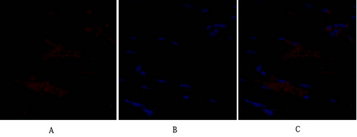
- Immunofluorescence analysis of rat-heart tissue. 1,SNAI 1 Polyclonal Antibody(red) was diluted at 1:200(4°C,overnight). 2, Cy3 labled Secondary antibody was diluted at 1:300(room temperature, 50min).3, Picture B: DAPI(blue) 10min. Picture A:Target. Picture B: DAPI. Picture C: merge of A+B
- Immunofluorescence analysis of rat-heart tissue. 1,SNAI 1 Polyclonal Antibody(red) was diluted at 1:200(4°C,overnight). 2, Cy3 labled Secondary antibody was diluted at 1:300(room temperature, 50min).3, Picture B: DAPI(blue) 10min. Picture A:Target. Picture B: DAPI. Picture C: merge of A+B

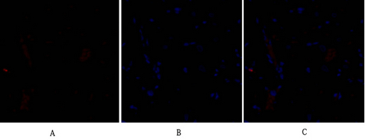
- Immunofluorescence analysis of rat-kidney tissue. 1,SNAI 1 Polyclonal Antibody(red) was diluted at 1:200(4°C,overnight). 2, Cy3 labled Secondary antibody was diluted at 1:300(room temperature, 50min).3, Picture B: DAPI(blue) 10min. Picture A:Target. Picture B: DAPI. Picture C: merge of A+B

- Immunofluorescence analysis of rat-kidney tissue. 1,SNAI 1 Polyclonal Antibody(red) was diluted at 1:200(4°C,overnight). 2, Cy3 labled Secondary antibody was diluted at 1:300(room temperature, 50min).3, Picture B: DAPI(blue) 10min. Picture A:Target. Picture B: DAPI. Picture C: merge of A+B

- Immunofluorescence analysis of rat-spleen tissue. 1,SNAI 1 Polyclonal Antibody(red) was diluted at 1:200(4°C,overnight). 2, Cy3 labled Secondary antibody was diluted at 1:300(room temperature, 50min).3, Picture B: DAPI(blue) 10min. Picture A:Target. Picture B: DAPI. Picture C: merge of A+B
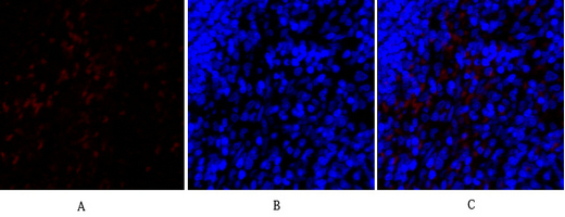
- Immunofluorescence analysis of rat-spleen tissue. 1,SNAI 1 Polyclonal Antibody(red) was diluted at 1:200(4°C,overnight). 2, Cy3 labled Secondary antibody was diluted at 1:300(room temperature, 50min).3, Picture B: DAPI(blue) 10min. Picture A:Target. Picture B: DAPI. Picture C: merge of A+B

- Western Blot analysis of various cells using SNAI 1 Polyclonal Antibody diluted at 1:1000 cells nucleus extracted by Minute TM Cytoplasmic and Nuclear Fractionation kit (SC-003,Inventbiotech,MN,USA).
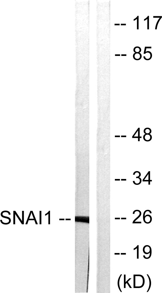
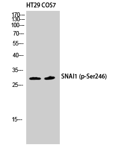
- Western blot analysis of lysates from HT29 cells, using SNAI1 Antibody. The lane on the right is blocked with the synthesized peptide.
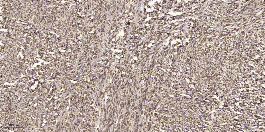
- Immunohistochemical analysis of paraffin-embedded human small intestinal carcinoma tissue. 1,primary Antibody was diluted at 1:200(4° overnight). 2, Sodium citrate pH 6.0 was used for antigen retrieval(>98°C,20min). 3,Secondary antibody was diluted at 1:200