LATS1/2 Polyclonal Antibody
- Catalog No.:YT2543
- Applications:WB;IHC;IF;ELISA
- Reactivity:Human;Mouse
- Target:
- LATS1/2
- Fields:
- >>Hippo signaling pathway;>>Hippo signaling pathway - multiple species
- Gene Name:
- LATS1/LATS2
- Protein Name:
- Serine/threonine-protein kinase LATS1/2
- Human Gene Id:
- 9113/26524
- Human Swiss Prot No:
- O95835/Q9NRM7
- Mouse Gene Id:
- 16798/50523
- Immunogen:
- The antiserum was produced against synthesized peptide derived from human LATS1/2. AA range:1041-1090
- Specificity:
- LATS1/2 Polyclonal Antibody detects endogenous levels of LATS1/2 protein.
- Formulation:
- Liquid in PBS containing 50% glycerol, 0.5% BSA and 0.02% sodium azide.
- Source:
- Polyclonal, Rabbit,IgG
- Dilution:
- WB 1:500 - 1:2000. IHC: 1:100-300 ELISA: 1:20000. IF 1:100-300 Not yet tested in other applications.
- Purification:
- The antibody was affinity-purified from rabbit antiserum by affinity-chromatography using epitope-specific immunogen.
- Concentration:
- 1 mg/ml
- Storage Stability:
- -15°C to -25°C/1 year(Do not lower than -25°C)
- Other Name:
- LATS1;WARTS;Serine/threonine-protein kinase LATS1;Large tumor suppressor homolog 1;WARTS protein kinase;h-warts;LATS2;KPM;Serine/threonine-protein kinase LATS2;Kinase phosphorylated during mitosis protein;Large tumor suppressor ho
- Observed Band(KD):
- 130-140kD
- Background:
- The protein encoded by this gene is a putative serine/threonine kinase that localizes to the mitotic apparatus and complexes with cell cycle controller CDC2 kinase in early mitosis. The protein is phosphorylated in a cell-cycle dependent manner, with late prophase phosphorylation remaining through metaphase. The N-terminal region of the protein binds CDC2 to form a complex showing reduced H1 histone kinase activity, indicating a role as a negative regulator of CDC2/cyclin A. In addition, the C-terminal kinase domain binds to its own N-terminal region, suggesting potential negative regulation through interference with complex formation via intramolecular binding. Biochemical and genetic data suggest a role as a tumor suppressor. This is supported by studies in knockout mice showing development of soft-tissue sarcomas, ovarian stromal cell tumors and a high sensitivity to carcinogenic treatmen
- Function:
- catalytic activity:ATP + a protein = ADP + a phosphoprotein.,cofactor:Magnesium.,function:Tumor suppressor which plays a critical role in maintenance of ploidy through its actions in both mitotic progression and the G1 tetraploidy checkpoint. Negatively regulates G2/M transition by down-regulating CDC2 kinase activity. Involved in the control of p53 expression. Affects cytokinesis by regulating actin polymerization through negative modulation of LIMK1. May also play a role in endocrine function.,PTM:Autophosphorylated and phosphorylated during M-phase of the cell cycle. Phosphorylated by STK3 at Ser-909 and Thr-1079, which results in its activation. Phosphorylated upon DNA damage, probably by ATM or ATR.,similarity:Belongs to the protein kinase superfamily. AGC Ser/Thr protein kinase family.,similarity:Contains 1 AGC-kinase C-terminal domain.,similarity:Contains 1 protein kinase domain.,
- Subcellular Location:
- Cytoplasm, cytoskeleton, microtubule organizing center, centrosome . Cytoplasm, cytoskeleton, spindle . Midbody . Cytoplasm, cytoskeleton, microtubule organizing center, spindle pole body . Localizes to the centrosomes throughout interphase but migrates to the mitotic apparatus, including spindle pole bodies, mitotic spindle, and midbody, during mitosis. .
- Expression:
- Expressed in all adult tissues examined except for lung and kidney.
RAS-association domain family 1A regulates the abnormal cell proliferation in psoriasis via inhibition of Yes-associated protein. JOURNAL OF CELLULAR AND MOLECULAR MEDICINE J Cell Mol Med. 2021 Jun;25(11):5070-5081 WB Human Psoriasis tissue, normal skin tissue
Yes-Associated Protein Expression is a Predictive Marker for Recurrence of Hepatocellular Carcinoma after Liver Transplantation. DIGESTIVE SURGERY 2015 Jan 22 WB Human HCC tissue
Jia, Jinjing, et al. "RAS‐association domain family 1A regulates the abnormal cell proliferation in psoriasis via inhibition of Yes‐associated protein." Journal of cellular and molecular medicine 25.11 (2021): 5070-5081.
Targeting the mechanism of IRF3 in sepsis-associated acute kidney injury via the Hippo pathway. Xingui Dai WB Human HK-2 cell
- June 19-2018
- WESTERN IMMUNOBLOTTING PROTOCOL
- June 19-2018
- IMMUNOHISTOCHEMISTRY-PARAFFIN PROTOCOL
- June 19-2018
- IMMUNOFLUORESCENCE PROTOCOL
- September 08-2020
- FLOW-CYTOMEYRT-PROTOCOL
- May 20-2022
- Cell-Based ELISA│解您多样本WB检测之困扰
- July 13-2018
- CELL-BASED-ELISA-PROTOCOL-FOR-ACETYL-PROTEIN
- July 13-2018
- CELL-BASED-ELISA-PROTOCOL-FOR-PHOSPHO-PROTEIN
- July 13-2018
- Antibody-FAQs
- Products Images

- Immunofluorescence analysis of A549. 1,primary Antibody(red) was diluted at 1:200(4°C overnight). 2, Goat Anti Rabbit IgG (H&L) - Alexa Fluor 594 Secondary antibody was diluted at 1:1000(room temperature, 50min).3, Picture B: DAPI(blue) 10min.

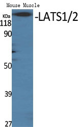
- Western Blot analysis of mouse-mscle cells using LATS1/2 Polyclonal Antibody diluted at 1:500

- Immunohistochemistry analysis of paraffin-embedded human breast carcinoma tissue, using LATS1/2 Antibody. The picture on the right is blocked with the synthesized peptide.
- Western blot analysis of LATS1/2 Antibody


