GIT1 Polyclonal Antibody
- Catalog No.:YT1908
- Applications:WB;IHC;IF;ELISA
- Reactivity:Human;Mouse;Rat
- Target:
- GIT1
- Fields:
- >>Endocytosis;>>Regulation of actin cytoskeleton;>>Epithelial cell signaling in Helicobacter pylori infection
- Gene Name:
- GIT1
- Protein Name:
- ARF GTPase-activating protein GIT1
- Human Gene Id:
- 28964
- Human Swiss Prot No:
- Q9Y2X7
- Mouse Gene Id:
- 216963
- Mouse Swiss Prot No:
- Q68FF6
- Rat Gene Id:
- 83709
- Rat Swiss Prot No:
- Q9Z272
- Immunogen:
- The antiserum was produced against synthesized peptide derived from human GIT1. AA range:561-610
- Specificity:
- GIT1 Polyclonal Antibody detects endogenous levels of GIT1 protein.
- Formulation:
- Liquid in PBS containing 50% glycerol, 0.5% BSA and 0.02% sodium azide.
- Source:
- Polyclonal, Rabbit,IgG
- Dilution:
- WB 1:500 - 1:2000. IHC 1:100 - 1:300. ELISA: 1:20000.. IF 1:50-200
- Purification:
- The antibody was affinity-purified from rabbit antiserum by affinity-chromatography using epitope-specific immunogen.
- Concentration:
- 1 mg/ml
- Storage Stability:
- -15°C to -25°C/1 year(Do not lower than -25°C)
- Other Name:
- GIT1;ARF GTPase-activating protein GIT1;ARF GAP GIT1;Cool-associated and tyrosine-phosphorylated protein 1;CAT-1;CAT1;G protein-coupled receptor kinase-interactor 1;GRK-interacting protein 1
- Observed Band(KD):
- 95kD
- Background:
- domain:The paxillin-binding domain is masked in the full-length protein and is regulated by ARHGEF6.,function:GTPase-activating protein for the ADP ribosylation factor family. May serve as a scaffold to bring together molecules to form signaling modules controlling vesicle trafficking, adhesion and cytoskeletal organization. Increases the speed of cell migration, as well as the size and rate of formation of protrusions, possibly by targeting PAK1 to adhesions and the leading edge of lamellipodia. Sequesters inactive non-tyrosine-phosphorylated paxillin in cytoplasmic complexes.,PTM:Phosphorylated on tyrosine residues by PTK2 and SRC in growing fibroblasts. Tyrosine-phosphorylation is increased following cell spreading on fibronectin, decreased in cells arrested in mitosis and increased in the ensuing G1 phase.,similarity:Contains 1 Arf-GAP domain.,similarity:Contains 3 ANK repeats.,subcellular location:Cycles between at least 3 distinct intracellular compartments, including focal adhesions, cytoplasmic complexes and membrane protrusions. During cell migration, when cells detach, moves from the adhesions into the cytoplasmic complexes towards the leading edge, while, when cells adhere, it is found in vinculin-containing adhesions. Recruitment to adhesions may be mediated by active tyrosine-phosphorylated paxillin.,subunit:Interacts with G protein-coupled receptor kinases: ADRBK1/GRK2, PPFIA1 and PPFIA4. Interacts with ARHGEF6/alpha-PIX, with ARHGEF7/beta-PIX, with PXN/paxillin and with PTK2/FAK (By similarity). Component of cytoplasmic complexes, which also contain PXN, ARHGEF6 and PAK1. Interacts with TGFB1I1.,
- Function:
- domain:The paxillin-binding domain is masked in the full-length protein and is regulated by ARHGEF6.,function:GTPase-activating protein for the ADP ribosylation factor family. May serve as a scaffold to bring together molecules to form signaling modules controlling vesicle trafficking, adhesion and cytoskeletal organization. Increases the speed of cell migration, as well as the size and rate of formation of protrusions, possibly by targeting PAK1 to adhesions and the leading edge of lamellipodia. Sequesters inactive non-tyrosine-phosphorylated paxillin in cytoplasmic complexes.,PTM:Phosphorylated on tyrosine residues by PTK2 and SRC in growing fibroblasts. Tyrosine-phosphorylation is increased following cell spreading on fibronectin, decreased in cells arrested in mitosis and increased in the ensuing G1 phase.,similarity:Contains 1 Arf-GAP domain.,similarity:Contains 3 ANK repeats.,subce
- Subcellular Location:
- Cytoplasm . Cell junction, synapse . Cell junction, synapse, presynapse . Cell junction, synapse, postsynapse . Cell junction, synapse, postsynaptic density . Cell junction, focal adhesion . Cell projection, lamellipodium . Cytoplasm, cytoskeleton, microtubule organizing center, centrosome . Cytoplasm, cytoskeleton, spindle pole . Cycles between at least 3 distinct intracellular compartments, including focal adhesions, cytosolic complexes, containing at least PXN/paxillin, ARHGEF7 and PAK1, and membrane protrusions. During cell migration, moves from the disassembling adhesions into the cytosol and towards the leading edge. In adherent cells, localizes to adhesions. Recruitment to adhesions may be mediated by RAC and active tyrosine-phosphorylated PXN (PubMed:11896197). May be present in bo
- Expression:
- Brain,Epithelium,Human testis,Liver,Lung,Melanoma,Muscle,Platelet,Skin,
- June 19-2018
- WESTERN IMMUNOBLOTTING PROTOCOL
- June 19-2018
- IMMUNOHISTOCHEMISTRY-PARAFFIN PROTOCOL
- June 19-2018
- IMMUNOFLUORESCENCE PROTOCOL
- September 08-2020
- FLOW-CYTOMEYRT-PROTOCOL
- May 20-2022
- Cell-Based ELISA│解您多样本WB检测之困扰
- July 13-2018
- CELL-BASED-ELISA-PROTOCOL-FOR-ACETYL-PROTEIN
- July 13-2018
- CELL-BASED-ELISA-PROTOCOL-FOR-PHOSPHO-PROTEIN
- July 13-2018
- Antibody-FAQs
- Products Images
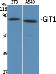
- Western Blot analysis of various cells using GIT1 Polyclonal Antibody
.jpg)
- Western Blot analysis of HepG2 cells using GIT1 Polyclonal Antibody
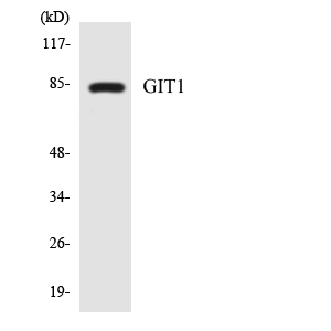
- Western blot analysis of the lysates from 293 cells using GIT1 antibody.
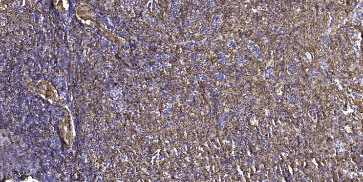
- Immunohistochemical analysis of paraffin-embedded human spleen tissue. 1,primary Antibody was diluted at 1:200(4° overnight). 2, Sodium citrate pH 6.0 was used for antigen retrieval(>98°C,20min). 3,Secondary antibody was diluted at 1:200


